

Acute sleep fragmentation does not alter pro-inflammatory cytokine gene expression in brain or peripheral tissues of leptin-deficient mice
- PMID: 29479505
- PMCID: PMC5822834
- DOI: 10.7717/peerj.4423
Acute sleep fragmentation does not alter pro-inflammatory cytokine gene expression in brain or peripheral tissues of leptin-deficient mice
Abstract
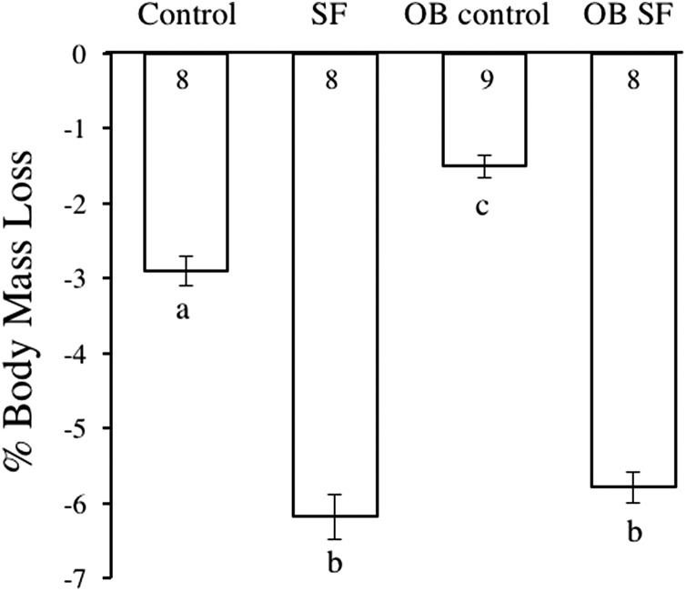
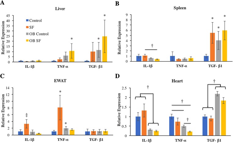
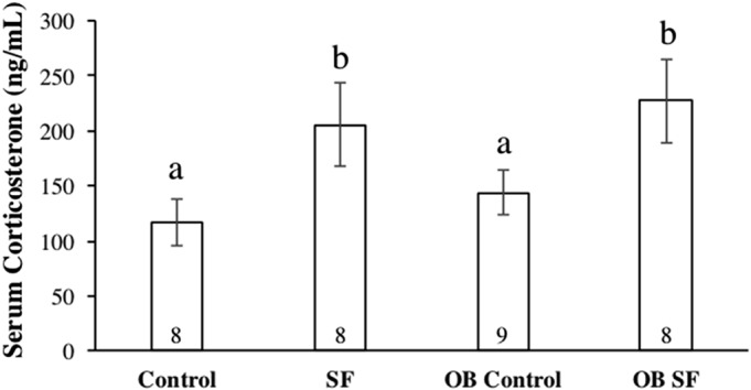
Obesity and sleep fragmentation (SF) are often co-occurring pro-inflammatory conditions in patients with obstructive sleep apnea. Leptin is a peptide hormone produced by adipocytes that has anorexigenic effects upon appetite while regulating immunity. The role of leptin in mediating inflammatory responses to SF is incompletely understood. Male C57BL/6j (lean) and ob/ob mice (leptin-deficient mice exhibiting obese phenotype) were subjected to SF or control conditions for 24 h using an automated SF chamber. Trunk blood and tissue samples from the periphery (liver, spleen, fat, and heart) and brain (hypothalamus, prefrontal cortex, and hippocampus) were collected. Quantitative PCR was used to determine relative cytokine gene expression of pro-inflammatory (IL-1β, TNF-α) and anti-inflammatory (TGF-β1) cytokines. Enzyme-linked immunosorbent assay (ELISA) was used to determine serum corticosterone concentration. Ob/ob mice exhibited elevated cytokine gene expression in liver (TNF-α, TGF-β1), heart (TGF-β1), fat (TNF-α), and brain (hippocampus, hypothalamus, prefrontal cortex: IL-1β, TNF-α) compared with wild-type mice. Conversely, leptin deficiency decreased pro-inflammatory cytokine gene expression in heart (IL-1β, TNF-α). SF significantly increased IL-1β and TNF-α gene expression in fat and TGF-β1 expression in spleen relative to controls, but only in wild-type mice. SF increased basal serum corticosterone regardless of genotype. Taken together, these findings suggest that leptin deficiency affects cytokine gene expression differently in the brain compared to peripheral tissues with minimal interaction from acute SF.
Keywords: Corticosterone; IL-1; IL-1β; Inflammation; Leptin; Ob/ob; Sleep fragmentation; Sleep loss; TGF-β1; TNF-α.
Conflict of interest statement
The authors declare that they have no competing interests.
Figures

References
-
- Ashley NT, Walton JC, Haim A, Zhang N, Prince LA, Fruchey AM, Lieberman RA, Weil ZM, Magalang UJ, Nelson RJ. Sleep deprivation attenuates endotoxin-induced cytokine gene expression independent of day length and circulating cortisol in male Siberian hamsters (Phodopus sungorus) Journal of Experimental Biology. 2013;216(14):2581–2586. doi: 10.1242/jeb.083832. - DOI - PubMed
Grants and funding
LinkOut - more resources
Full Text Sources
Other Literature Sources
Miscellaneous
