

Review
doi: 10.4103/ijo.IJO_831_17.
Dynamic corneal deformation response and integrated corneal tomography
Affiliations
- PMID: 29480246
- PMCID: PMC5859590
- DOI: 10.4103/ijo.IJO_831_17
Item in Clipboard
Review
Dynamic corneal deformation response and integrated corneal tomography
Indian J Ophthalmol.
2018 Mar.
Abstract
Measuring corneal biomechanical properties is still challenging. There are several clinical applications for biomechanical measurements, including the detection of mild or early forms of ectatic corneal diseases. This article reviews clinical applications for biomechanical measurements provided by the Corvis ST dynamic non contact tonometer.
Keywords: Corneal biomechanics; corneal tomography; ectasia; keratoconus.
Conflict of interest statement
There are no conflicts of interest.
Figures
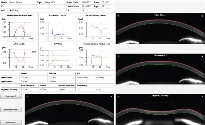
Corvis ST overview display
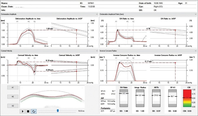
Corvis ST corneal biomechanical index display
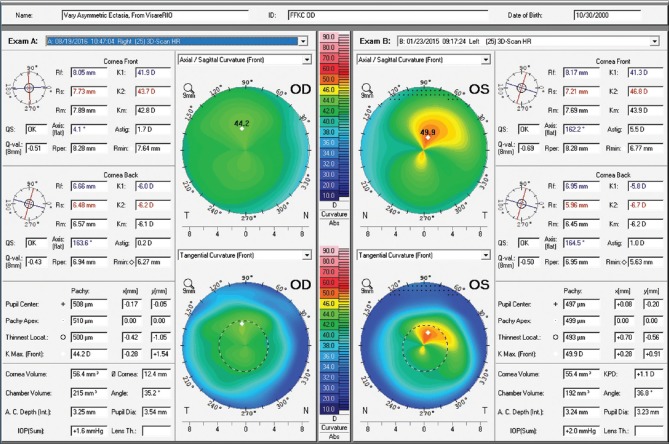
Axial and tangential curvature maps of the front corneal surface from both eyes, obtained with the Pentacam HR corneal tomography (Oculus Optikgeräte GmbH, Wetzlar, Germany). Very mild asymmetry is observed in OD and advanced KC in OS

Anterior surface corneal topography from OD at the first visit in 2015 (C) and with 2 years of follow up in 2017 (A). Anterior surface corneal topography from OS in pre (D) and post operative (B) periods
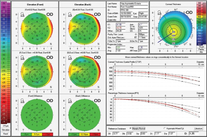
Belin-Ambrósio Enhanced Ectasia Display from the Pentacam of OD. Note ARTmax value of 371 and Belin/Ambrósio Deviation of 1.25
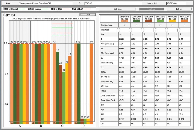
Figur 6: D-The Belin ABCD keratoconus staging
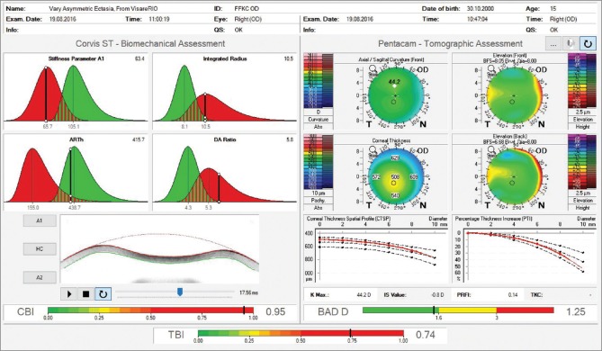
Ambrósio, Robers and Vinciguerra tomographic/biomechanical index display. Observe an abnormal corneal biomechanical index value of 0.95 and abnormal tomographic/biomechanical index value of 0.74

Axial curvature maps of the front corneal surface obtained with the Keratograph 5M (Oculus Optikgeräte GmbH, Wetzlar, Germany). We can observe a truncated bow tie and skewed radial axis pattern in OD, but innocent findings in OS. The bottom part of the figure presents Pentacam maps with Belin ABCD keratoconus staging
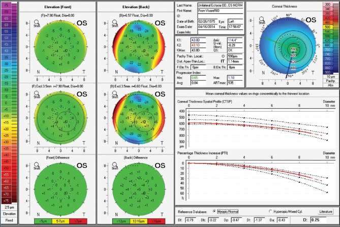
Belin-Ambrósio Enhanced Ectasia Display from the Pentacam of OS. Unremarkable findings are observed
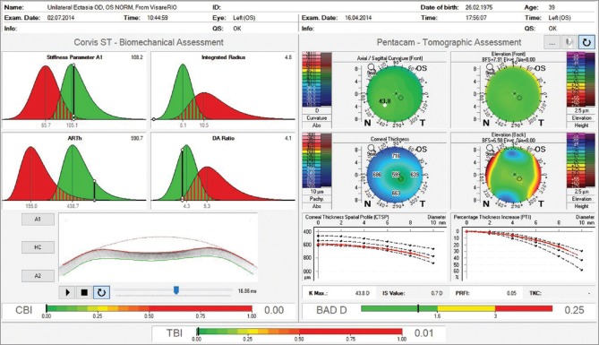
Pentacam/Corvis ST Ambrósio, Roberts and Vinciguerra tomographic/biomechanical index display
Similar articles
-
Long-term Evaluation of Corneal Biomechanical Properties After Corneal Cross-linking for Keratoconus: A 4-Year Longitudinal Study.J Refract Surg. 2018 Dec 1;34(12):849-856. doi: 10.3928/1081597X-20181012-02. J Refract Surg. 2018. PMID: 30540368
-
Variability of Corneal Deformation Response in Normal and Keratoconic Eyes.Optom Vis Sci. 2015 Jul;92(7):e149-53. doi: 10.1097/OPX.0000000000000628. Optom Vis Sci. 2015. PMID: 26002009
-
Assessment of corneal biomechanical parameters in healthy and keratoconic eyes using dynamic bidirectional applanation device and dynamic Scheimpflug analyzer.J Cataract Refract Surg. 2019 Jun;45(6):778-788. doi: 10.1016/j.jcrs.2018.12.015. Epub 2019 Mar 20. J Cataract Refract Surg. 2019. PMID: 30902432
-
Corneal biomechanics in patients with allergic conjunctivitis.Indian J Ophthalmol. 2024 Nov 1;72(Suppl 5):S728-S733. doi: 10.4103/IJO.IJO_654_24. Epub 2024 Sep 19. Indian J Ophthalmol. 2024. PMID: 39297487 Free PMC article. Review.
-
Biomechanical Diagnostics of the Cornea.Int Ophthalmol Clin. 2017 Summer;57(3):75-86. doi: 10.1097/IIO.0000000000000172. Int Ophthalmol Clin. 2017. PMID: 28590282 Free PMC article. Review.
Cited by
-
Keratoconus and Corneal Ectasia with Relatively Low Keratometry.Ophthalmol Ther. 2024 Jul;13(7):2023-2035. doi: 10.1007/s40123-024-00964-5. Epub 2024 Jun 2. Ophthalmol Ther. 2024. PMID: 38824471 Free PMC article.
-
Assessment of corneal topographic, tomographic, densitometric, and biomechanical properties of Fabry patients with ocular manifestations.Graefes Arch Clin Exp Ophthalmol. 2020 May;258(5):1057-1064. doi: 10.1007/s00417-019-04593-8. Epub 2020 Jan 8. Graefes Arch Clin Exp Ophthalmol. 2020. PMID: 31915973
-
A Critical Assessment of Friedenwald's Technique for Estimating the Coefficient of Rigidity of the Cornea.J Ophthalmol. 2022 Oct 4;2022:6775064. doi: 10.1155/2022/6775064. eCollection 2022. J Ophthalmol. 2022. PMID: 36237558 Free PMC article.
-
Early elastic and viscoelastic corneal biomechanical changes after photorefractive keratectomy and small incision lenticule extraction.Int Ophthalmol. 2024 Jul 2;44(1):302. doi: 10.1007/s10792-024-03169-8. Int Ophthalmol. 2024. PMID: 38954134
-
Biomechanical diagnostics of the cornea.Eye Vis (Lond). 2020 Feb 5;7:9. doi: 10.1186/s40662-020-0174-x. eCollection 2020. Eye Vis (Lond). 2020. PMID: 32042837 Free PMC article. Review.
References
-
- Andreassen TT, Simonsen AH, Oxlund H. Biomechanical properties of keratoconus and normal corneas. Exp Eye Res. 1980;31:435–41. - PubMed
-
- Liu J, Roberts CJ. Influence of corneal biomechanical properties on intraocular pressure measurement: Quantitative analysis. J Cataract Refract Surg. 2005;31:146–55. - PubMed
-
- Dupps WJ., Jr Biomechanical modeling of corneal ectasia. J Refract Surg. 2005;21:186–90. - PubMed
-
- Luce DA. Determining in vivo biomechanical properties of the cornea with an ocular response analyzer. J Cataract Refract Surg. 2005;31:156–62. - PubMed
Publication types
MeSH terms
LinkOut - more resources
Full Text Sources
