. 2018 Mar;66(3):433-438.
doi: 10.4103/ijo.IJO_845_17.

Spectral domain optical coherence tomography evaluation of macular changes in Eales disease

Affiliations

Spectral domain optical coherence tomography evaluation of macular changes in Eales disease

Neha Goel et al. Indian J Ophthalmol. 2018 Mar.

Abstract

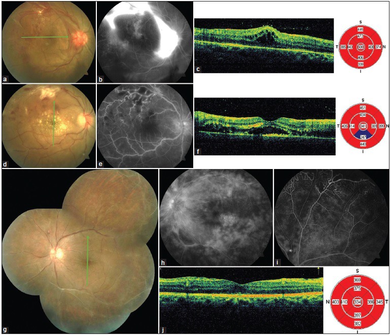
Purpose: The purpose of the study was to describe macular changes in treatment-naïve eyes with Eales disease using spectral domain optical coherence tomography (SD-OCT).

Methods: A cross-sectional study was done on 79 eyes of 66 patients with Eales disease. Best-corrected visual acuity (BCVA), slit-lamp biomicroscopy (SLB), indirect ophthalmoscopy, fundus fluorescein angiography (FFA), and quantitative (central macular thickness [CMT]) and qualitative analysis on SD-OCT were performed.

Results: Forty-six (58.2%) eyes had macular involvement as assessed with SD-OCT, while in 33 (41.8%) eyes, macula was not affected. Macular edema was the most common feature when macula was affected followed by epiretinal membrane. Mean CMT was higher (315.3 ± 102.3 μm) in eyes with macular involvement than those without it (243.8 ± 19.3 μm). Eyes with active vasculitis involving larger vessels and neovascularization had greater chance of macular involvement. SLB and FFA alone missed 28.3% and 50% eyes with macular abnormalities on SD-OCT, respectively.

Conclusion: While the clinical description of Eales disease points mainly to a peripheral location, macular involvement can be commonly picked up when SD-OCT is used. Macular involvement when present is associated with a poorer BCVA.

Keywords: Eales disease; epiretinal membrane; macula; macular edema; retinal vasculitis; spectral domain optical coherence tomography.


Conflict of interest statement

There are no conflicts of interest.

Figures

Figure 1
Figure 1
Distribution of eyes with and without macular involvement according to the stage of Eales disease given by Saxena et al.:[4] Stage 1 – periphlebitis of small (1A) and large (1B) caliber vessels, Stage 2A – capillary nonperfusion, 2B – neovascularization elsewhere/of the disc, Stage 3A – fibrovascular proliferation, 3B – vitreous hemorrhage
Figure 2
Figure 2
(a) Fundus photograph and (b and c) fluorescein angiogram of a 28 years male with Eales disease Stage 3B and best-corrected visual acuity 0.67 showing peripheral neovascularization, capillary nonperfusion, and a normal macula (d) spectral domain optical coherence tomography revealed epiretinal membrane inferiorly, in addition (e) fundus photograph and (f and g) fluorescein angiogram of a 30-year-old male with best-corrected visual acuity 0.05 showing superotemporal healed vasculitis, peripheral collaterals, and a normal macula (h) spectral domain optical coherence tomography showed temporal macular thinning not visible clinically or on fluorescein angiogram
Figure 3
Figure 3
(a) Fundus photograph and (b) fluorescein angiogram of a 22-year-old male with Eales disease Stage 3A and best-corrected visual acuity 0.1 showing fibrovascular proliferation at the disc (c) spectral domain optical coherence tomography showed cystoid macular edema (d) fundus photograph and (e) fluorescein angiogram of a 25-year-old male with best-corrected visual acuity 0.05 showing superotemporal periphlebitis and macular hard exudates (Eales disease Stage 1B) (f) spectral domain optical coherence tomography showed macular edema and subfoveal neurosensory detachment (g) fundus photograph of a 30-year-old male showing active vasculitis in all quadrants (Eales disease Stage 2B). Best-corrected visual acuity was 0.167. Fluorescein angiogram showed diffuse vascular leakage (h), and superior collaterals (i), (j) spectral domain optical coherence tomography showed epiretinal membrane and macular edema
Figure 4
Figure 4
(a) Fundus photograph of a 17-year-old male showing an inferior sclerosed vessel with an altered foveal reflex. Best-corrected visual acuity was 0.67. Fluorescein angiogram showed inferior macular nonperfusion (b) with inferonasal collaterals (c). This was Stage 2A of Eales disease (d) spectral domain optical coherence tomography demonstrated gross macular thinning. (e) Fundus photograph of a 37-year-old male with Eales disease Stage 2B and best-corrected visual acuity 0.1 showing fibrous proliferation at the disc and a macular hole. Fluorescein angiogram showed staining at the disc and window defects corresponding to the macular hole (f) with neovascularization inferiorly (g), (h) spectral domain optical coherence tomography confirmed a full-thickness macular hole
Figure 5
Figure 5
A 39-year-old male presented with sudden loss of vision in his left eye for 2 days. Best-corrected visual acuity was 0.016. (a) Fundus showed a premacular hemorrhage giving the appearance of a “double ring” (outer black and inner white arrows) with periphlebitis of the inferotemporal vein. (b) Fluorescein angiogram showed blocked fluorescence in the area of the hemorrhage with inferior active vasculitis. (c) Spectral domain optical coherence tomography demonstrated that the outer ring was that of a subinternal limiting membrane hemorrhage and the inner ring was composed of hyperreflective deposits below the internal limiting membrane causing shadowing of the remaining retinal layers

Comment in

  • Macular changes in retinal vasculitis.
    Saurabh K, Roy R. Saurabh K, et al. Indian J Ophthalmol. 2018 Mar;66(3):439. doi: 10.4103/ijo.IJO_1232_17. Indian J Ophthalmol. 2018. PMID: 29480259 Free PMC article. No abstract available.

References

    1. Das T, Pathengay A, Hussain N, Biswas J. Eales’ disease: Diagnosis and management. Eye (Lond) 2010;24:472–82. - PubMed
    1. Biswas J, Sharma T, Gopal L, Madhavan HN, Sulochana KN, Ramakrishnan S, et al. Eales disease – An update. Surv Ophthalmol. 2002;47:197–214. - PubMed
    1. Badrinath SS, Honnatti MR. Vitrectomy in Eales disease. Acta Int Cong Ophthalmol. 1982;24:536.
    1. Saxena S, Kumar D. New classification system-based visual outcome in Eales’ disease. Indian J Ophthalmol. 2007;55:267–9. - PubMed
    1. Saxena S, Kumar D. Macular involvement in Eales disease. Ann Ophthalmol. 2000;32:98–100.

Supplementary concepts