

Subretinal tissue plasminogen-assisted vitrectomy for posttraumatic full-thickness macular hole with submacular hemorrhage
- PMID: 29480275
- PMCID: PMC5859619
- DOI: 10.4103/ijo.IJO_815_17
Subretinal tissue plasminogen-assisted vitrectomy for posttraumatic full-thickness macular hole with submacular hemorrhage
Abstract
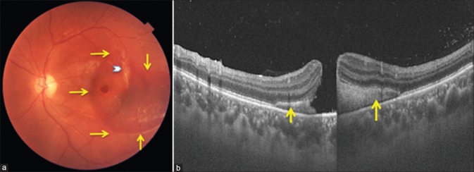
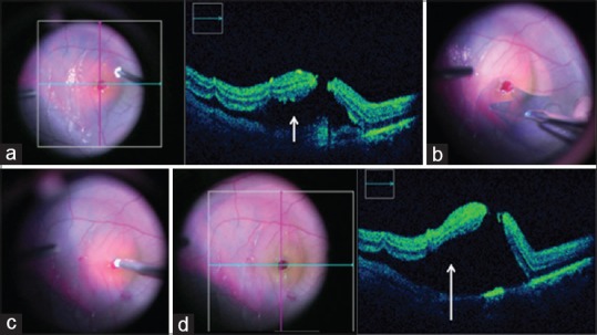
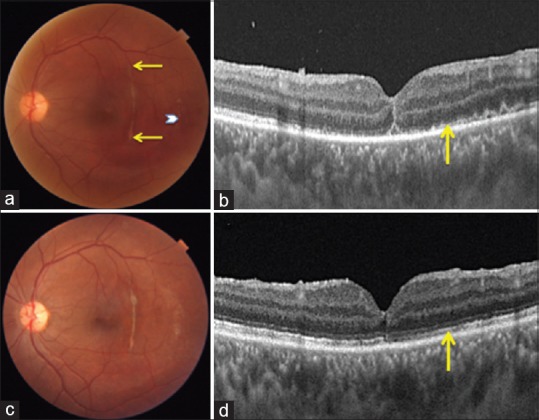
A young male presented with diminution of vision left eye, attributable to full-thickness macular hole, and submacular hemorrhage, following closed globe injury 2 weeks ago. The patient was managed successfully with 25-gauge vitrectomy, subretinal injection of tissue plasminogen activator and aspiration of liquefied blood through the macular hole, internal limiting membrane peeling, short-acting gas tamponade, and prone positioning. This resulted in good visual improvement, type 1 macular hole closure, and restoration of foveal architecture. The outcome and rationale of treatment in this unique scenario is discussed.
Keywords: Closed globe injury; full-thickness macular hole; pars plana vitrectomy; subretinal hemorrhage; subretinal tissue plasminogen activator.
Conflict of interest statement
There are no conflicts of interest.
Figures
Similar articles
-
Successful displacement of a traumatic submacular hemorrhage in a 13-year-old boy treated by vitrectomy, subretinal injection of tissue plasminogen activator and intravitreal air tamponade: a case report.BMC Ophthalmol. 2015 Aug 7;15:94. doi: 10.1186/s12886-015-0090-3. BMC Ophthalmol. 2015. PMID: 26250101 Free PMC article.
-
Subretinal injection of recombinant tissue plasminogen activator for submacular hemorrhage associated with ruptured retinal arterial macroaneurysm.Graefes Arch Clin Exp Ophthalmol. 2015 Oct;253(10):1663-9. doi: 10.1007/s00417-014-2861-6. Epub 2014 Nov 25. Graefes Arch Clin Exp Ophthalmol. 2015. PMID: 25418034
-
Management of macular hole and submacular hemorrhage in the same eye.Graefes Arch Clin Exp Ophthalmol. 2007 Apr;245(4):609-11. doi: 10.1007/s00417-006-0349-8. Epub 2006 Jul 27. Graefes Arch Clin Exp Ophthalmol. 2007. PMID: 16871381
-
The Role of Subretinal Injection in Ophthalmic Surgery: Therapeutic Agent Delivery and Other Indications.Int J Mol Sci. 2023 Jun 23;24(13):10535. doi: 10.3390/ijms241310535. Int J Mol Sci. 2023. PMID: 37445711 Free PMC article. Review.
-
Treatment for submacular hemorrhage associated with neovascular age-related macular degeneration.Semin Ophthalmol. 2011 Nov;26(6):361-71. doi: 10.3109/08820538.2011.585368. Semin Ophthalmol. 2011. PMID: 22044334 Review.
References
-
- Miller JB, Yonekawa Y, Eliott D, Kim IK, Kim LA, Loewenstein JI, et al. Long-term follow-up and outcomes in traumatic macular holes. Am J Ophthalmol. 2015;160:1255–80. - PubMed
-
- Berrocal MH, Lewis ML, Flynn HW., Jr Variations in the clinical course of submacular hemorrhage. Am J Ophthalmol. 1996;122:486–93. - PubMed
-
- Hochman MA, Seery CM, Zarbin MA. Pathophysiology and management of subretinal hemorrhage. Surv Ophthalmol. 1997;42:195–213. - PubMed
-
- Benner JD, Hay A, Landers MB, 3rd, Hjelmeland LM, Morse LS. Fibrinolytic-assisted removal of experimental subretinal hemorrhage within seven days reduces outer retinal degeneration. Ophthalmology. 1994;101:672–81. - PubMed
Publication types
MeSH terms
Substances
LinkOut - more resources
Full Text Sources
Medical
Miscellaneous
