. 2018 May 4;57(19):5257-5261.
doi: 10.1002/anie.201711825. Epub 2018 Mar 26.

High-Throughput Kinetic Analysis for Target-Directed Covalent Ligand Discovery

Affiliations

High-Throughput Kinetic Analysis for Target-Directed Covalent Ligand Discovery

Gregory B Craven et al. Angew Chem Int Ed Engl. .

Abstract

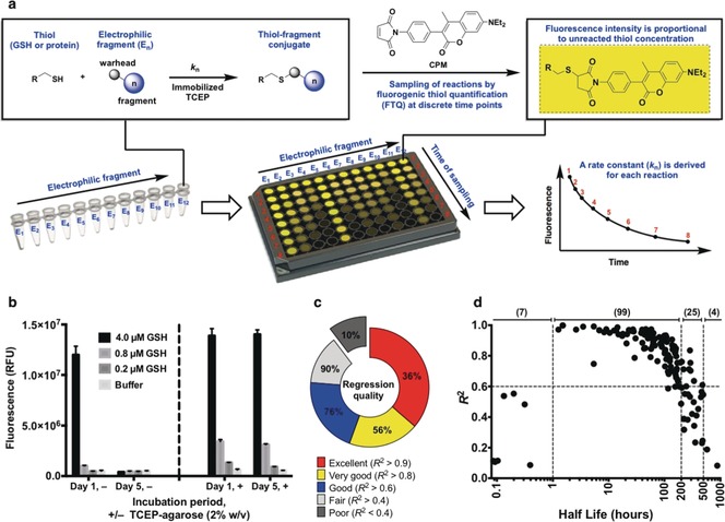
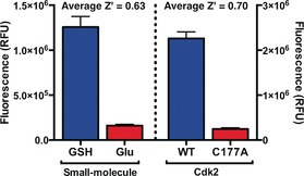
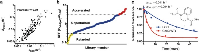
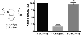
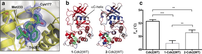
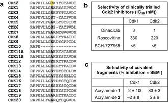
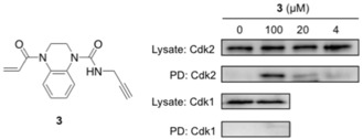
Cysteine-reactive small molecules are used as chemical probes of biological systems and as medicines. Identifying high-quality covalent ligands requires comprehensive kinetic analysis to distinguish selective binders from pan-reactive compounds. Quantitative irreversible tethering (qIT), a general method for screening cysteine-reactive small molecules based upon the maximization of kinetic selectivity, is described. This method was applied prospectively to discover covalent fragments that target the clinically important cell cycle regulator Cdk2. Crystal structures of the inhibitor complexes validate the approach and guide further optimization. The power of this technique is highlighted by the identification of a Cdk2-selective allosteric (type IV) kinase inhibitor whose novel mode-of-action could be exploited therapeutically.

Keywords: Cdk2; covalent inhibition; fragment-based drug discovery; kinetics; protein modification.


Conflict of interest statement

G.B.C., S.M., A.A., and D.J.M. are co‐inventors on a patent application covering qIT: PCT/GB2017/052456.

Figures

Figure 1
Figure 1
Quantitative irreversible tethering (qIT). a) Assay overview. The target thiol (5 μm) is reacted with electrophilic fragments (0.5 mm) under pseudo‐first‐order conditions in the presence of TCEP‐agarose (2 % w/v). Reaction progress is followed by discrete measurements of residual target thiol concentration using the fluorogenic probe CPM and rate constants are derived from exponential regression analysis. b) TCEP‐agarose prevents aerobic thiol oxidation. Glutathione solutions were stored ±TCEP‐agarose (2 % w/v). Agarose‐beads were separated by centrifugation prior to fluorogenic thiol quantification with CPM after one hour or five days. c) Performance of qIT in determining rate constants for library members in reaction with glutathione is reflected by the coefficient of determination (R 2) for each exponential regression. d) Accuracy of kinetic modeling as a function of reaction half‐life (t 1/2). Optimum assay performance is achieved when 1>t 1/2>200 hours.
Figure 2
Figure 2
Z′ factor analysis for qIT. Positives=Cdk2(WT) and GSH; negatives=Cdk2(C177A) and glutamate (n=72).
Figure 3
Figure 3
a) Correlation analysis comparing fragment reactivity with glutathione (k GSH) to average fragment reactivity across seven Cdk2 mutants ( protein). b) Distribution of rate enhancement factors for the covalent fragment library screened against Cdk2(WT). c) DMSO‐Normalized fluorescence data from qIT assay for acrylamide 1 (0.5 mm) in reaction with Cdk2(WT) or glutathione (5 μm) (n=2).
Figure 4
Figure 4
Kinase activity of 1‐Cdk2(WT) (16.8±3.1 %) and 2‐Cdk2(WT) (96.6±5.6 %) are reported relative to Cdk2(WT) (n=3; error bars and ± denote SEM).
Figure 5
Figure 5
a) Crystal structure of 1‐Cdk2(WT) (resolution: 1.83 Å, PDB ID: 5OSJ). 2 F oF c Electron density map (blue) is contoured at 1σ around C177 (yellow) and the ligand (green). b) Molecular dynamics simulations (50 ns) of 1‐Cdk2(WT) and 2‐Cdk2(WT) (resolution: 1.72 Å, PDB ID: 5OO0). Atomic flexibility (root mean square fluctuations) was compared to a similar simulation for Cdk2(WT) (ligands in green, flexibility: red=increase; blue=decrease and grey=unperturbed). c) T m were determined by TdCD (n=3; error bars=SD). **, *** and **** denote P<0.01, 0.001 and 0.0001 respectively in two‐tailed T‐test).
Figure 6
Figure 6
a) Sequence alignment of Cdk family. Cys177 is highlighted in yellow. b) Published selectivity profiles of selected Cdk2 inhibitors.29 c) Active Cdk1/Cdk2:cyclin A2 was incubated with acrylamides 1 and 2 (0.5 mm) for 24 hours and then their in vitro kinase activity was measured (n=3).
Figure 7
Figure 7
HeLa cells were treated with 3 for 6 hours. After tagging with biotin‐azide, Neutavidin pulldown was performed. Western blot analysis (anti‐Cdk2 and Cdk1) was performed before (Lysate) and after pulldown (PD).

References

    1. Surade S., Blundell T. L., Chem. Biol. 2012, 19, 42–50. - PubMed
    1. Scott D. E., Coyne A. G., Hudson S. A., Abell C., Biochemistry 2012, 51, 4990–5003. - PubMed
    1. Erlanson D. A., Wells J. A., Braisted A. C., Annu. Rev. Biophys. Biomol. Struct. 2004, 33, 199–223. - PubMed
    1. Kathman S. G., Span I., Smith A. T., Xu Z., Zhan J., Rosenzweig A. C., Statsyuk A. V., J. Am. Chem. Soc. 2015, 137, 12442–12445. - PMC - PubMed
    1. Krishnan S., Miller R. M., Tian B., Mullins R. D., Jacobson M. P., Taunton J., J. Am. Chem. Soc. 2014, 136, 12624–12630. - PMC - PubMed

Publication types

MeSH terms