. 2018 Mar 21;63(6):064002.
doi: 10.1088/1361-6560/aab241.

In vivo optoacoustic temperature imaging for image-guided cryotherapy of prostate cancer

Affiliations

In vivo optoacoustic temperature imaging for image-guided cryotherapy of prostate cancer

E V Petrova et al. Phys Med Biol. .

Abstract

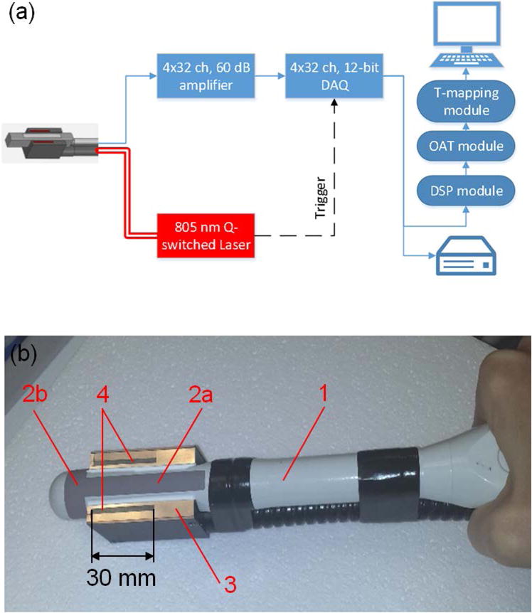
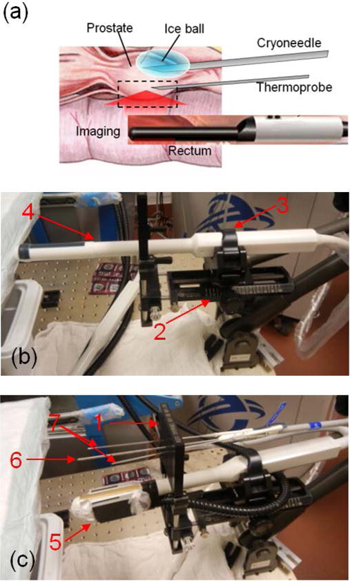
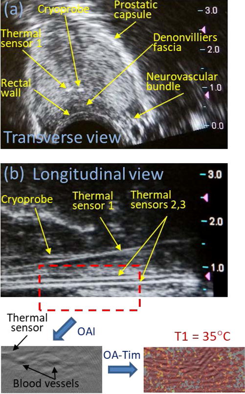
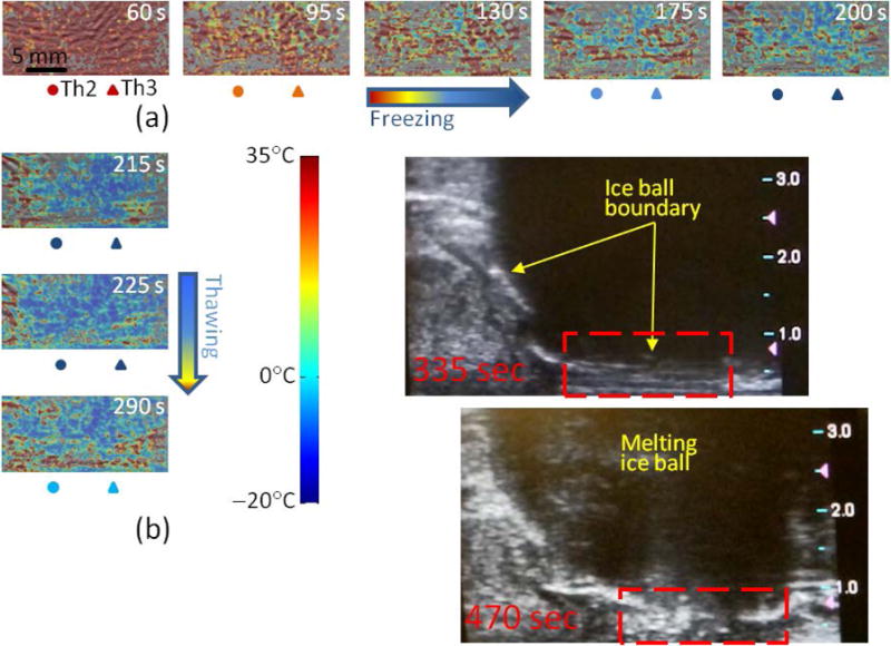
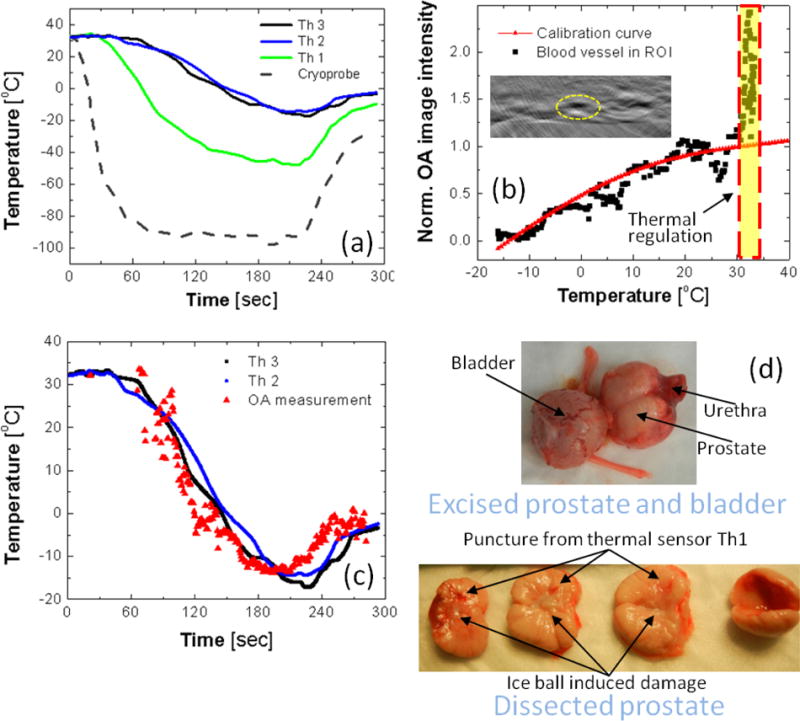
The objective of this study is to demonstrate in vivo the feasibility of optoacoustic temperature imaging during cryotherapy of prostate cancer. We developed a preclinical prototype optoacoustic temperature imager that included pulsed optical excitation at a wavelength of 805 nm, a modified clinical transrectal ultrasound probe, a parallel data acquisition system, image processing and visualization software. Cryotherapy of a canine prostate was performed in vivo using a commercial clinical system, Cryocare® CS, with an integrated ultrasound imaging. The universal temperature-dependent optoacoustic response of blood was employed to convert reconstructed optoacoustic images to temperature maps. Optoacoustic imaging of temperature during prostate cryotherapy was performed in the longitudinal view over a region of 30 mm (long) × 10 mm (deep) that covered the rectum, the Denonvilliers fascia, and the posterior portion of the treated gland. The transrectal optoacoustic images showed high-contrast vascularized regions, which were used for quantitative estimation of local temperature profiles. The constructed temperature maps and their temporal dynamics were consistent with the arrangement of the cryoprobe and readouts of the thermal needle sensors. The temporal profiles of the readouts from the thermal needle sensors and the temporal profile estimated from the normalized optoacoustic intensity of the selected vascularized region showed significant resemblance, except for the initial overshoot, that may be explained as a result of the physiological thermoregulatory compensation. The temperature was mapped with errors not exceeding ±2 °C (standard deviation) consistent with the clinical requirements for monitoring cryotherapy of the prostate. In vivo results showed that the optoacoustic temperature imaging is a promising non-invasive technique for real-time imaging of tissue temperature during cryotherapy of prostate cancer, which can be combined with transrectal ultrasound-the current standard for guiding clinical cryotherapy procedure.


Figures

Figure 1
Figure 1
Biological effects of cryotherapy.
Figure 2
Figure 2
(a) Schematic of the optoacoustic thermometry prototype for monitoring cryotherapy of prostate. DAQ – data acquisition, DSP – digital signal processing, OAT – optoacoustic tomography, T-mapping – temperature imaging. (b) Transrectal ultrasound (TRUS) probe (1) incorporates two orthogonal arrays of transducers: longitudinal (2a) and transverse (2b); the output terminal of the fiberoptic bundle (3) enables longitudinal optoacoustic view of the prostate excited by 805-nm laser pulses through the light-emitting apertures (4).
Figure 3
Figure 3
(a) Schematic showing in vivo optoacoustic temperature monitoring during cryotherapy of the canine prostate. Photographs show settings with transrectal probes for ultrasound imaging (b) and optoacoustic thermometry (c). 1 – CryoGrid™ fixture; 2 – probe holder; 3 – quick-latch; 4 – transrectal ultrasound (TRUS) probe; 5 – optoacoustic thermometry probe; 6 – cryoneedle; 7 – thermal sensors (thermoprobes).
Figure 4
Figure 4
(a) Transverse and (b) longitudinal ultrasound images of a dog’s prostate prior to initiation of cryotherapy. Red dashed rectangle on the panel (b) indicates the area that included rectal wall and Denonvilliers fascia, which was used for construction of optoacoustic temperature maps. OAI – optoacoustic imaging; OA-Tim – optoacoustic temperature imaging; T1 – initial temperature of the monitored prostate tissue.
Figure 5
Figure 5
In vivo optoacoustic imaging of temperature in the area near the rectal wall and Denonvilliers fascia during the freezing (a) and passive thawing (b) cycles of prostate cryotherapy. The timestamp indicates the moment of the frame’s acquisition after the initiation of the freezing cycle. The temperatures logged by the thermal sensors Th2 and Th3 set in the Denonvilliers fascia are indicated for each frame in the color code according to the shown color palette. Panel (b) also shows longitudinal ultrasound images of the dog’s prostate immediately after the cryotherapy. Ice ball is clearly visible as a large hypoechoic mass. Red dashed rectangle indicates the optoacoustically monitored region of interest.
Figure 6
Figure 6
(a) The temporal temperature profiles recorded by a thermocouple inside the cryoprobe and by three thermal sensors set inside (Th1) and nearby (Th2 and Th3) the prostate treated with cryotherapy. (b) Normalized optoacoustic image intensity of the high-contrast object (blood vessel) inside the region of interest (ROI) shown on the inset as a function of average temperature recorded by nearby thermal sensors Th2 and Th3; For comparison, the in vitro obtained universal calibration curve of blood is shown by the red solid line. (c) The temporal temperature profile reconstructed for the object in ROI shown on the panel (b); for comparison, the temporal temperature profiles logged by the nearby thermal sensors Th2 and Th3 are shown by the black and blue solid lines. (d) The dog’s prostate was excised and dissected following the cryotherapy to show transverse sections across the urethra demonstrating damage induced by the freezing.

References

    1. Agarwal PK, Sadetsky N, Konety BR, Resnick MI, Carroll PR, Cancer of the Prostate Strategic Urological Research E Treatment failure after primary and salvage therapy for prostate cancer: likelihood, patterns of care, and outcomes. Cancer. 2008;112:307–14. - PubMed
    1. ANSI. Americal National Standard for Safe Use of Lasers. Americal National Standards Institutes. 2007;Z136.1:6–24.
    1. Bahmani B, Bacon D, Anvari B. Erythrocyte-derived photo-theranostic agents: hybrid nano-vesicles containing indocyanine green for near-infrared imaging and therapeutic applications. Sci Rep. 2013;3:2180. - PMC - PubMed
    1. Bjerklund Johansen TE, Witzsch U, Greene D. Salvage treatment in prostate cancer: a clinical approach. Expert review of anticancer therapy. 2013;13:613–23. - PubMed
    1. Chen YS, Frey W, Aglyamov S, Emelianov S. Environment-dependent generation of photoacoustic waves from plasmonic nanoparticles. Small (Weinheim an der Bergstrasse, Germany) 2012;8:47–52. - PMC - PubMed

Publication types