

Congenital left atrial appendage aneurysm: A rare case report and literature review
- PMID: 29480827
- PMCID: PMC5943883
- DOI: 10.1097/MD.0000000000009344
Congenital left atrial appendage aneurysm: A rare case report and literature review
Abstract
Rationale: Left atrial appendage aneurysms (LAAA) are rare. Patients with LAAA are often diagnosed incidentally or after cardiac tachyarrhythmia or systemic thromboembolism happen. Early diagnosis and surgical resection is of utmost importance to prevent hazardous adverse events.
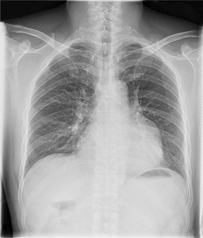
Patient concerns: We present a case of 46-year-old man with congenital LAAA. The individual in this manuscript has given written informed consent to publish these case details.
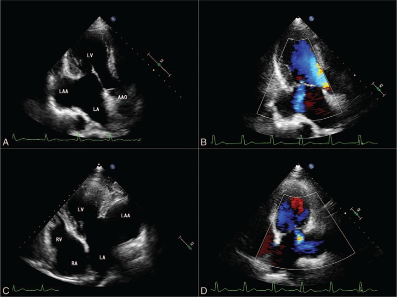
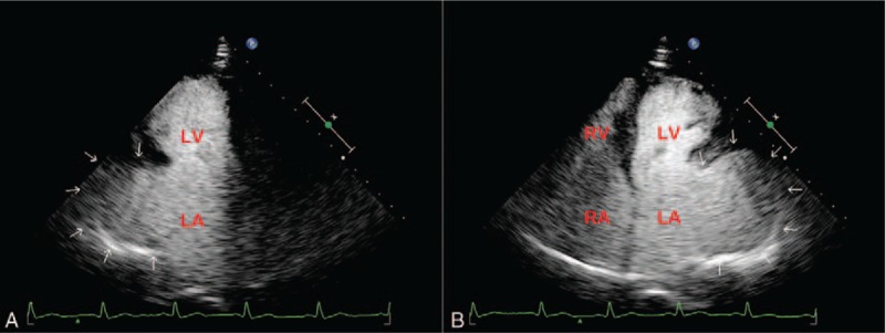
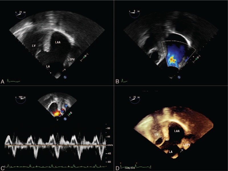
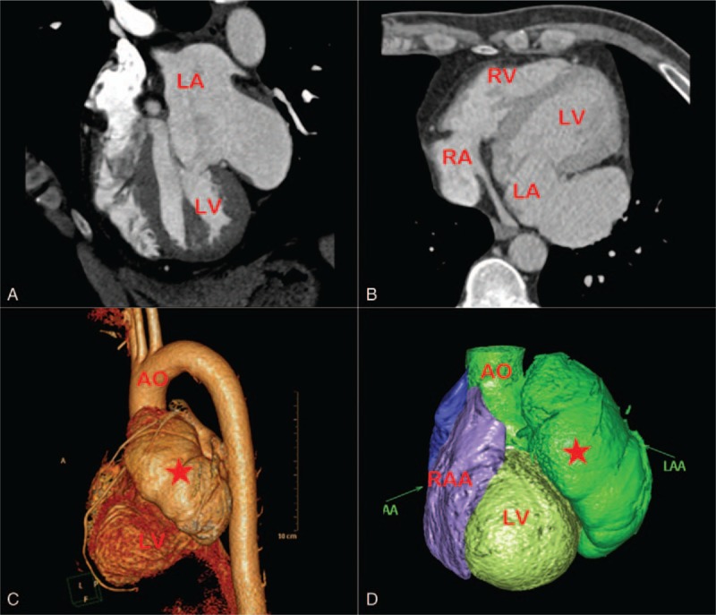
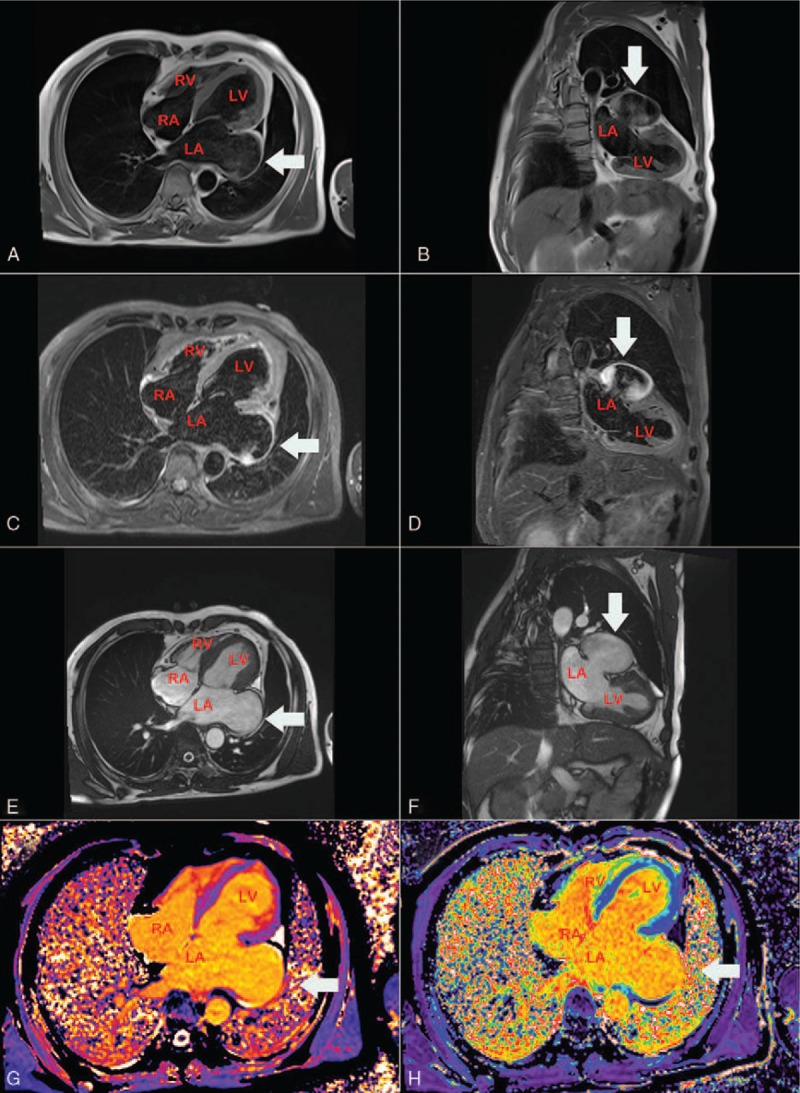
Diagnoses: Imaging studies, such as echocardiography, cardiovascular computed tomography (CT) and magnetic resonance imaging (MRI), demonstrated the large cavity arising from the left atrial appendage. The diagnosis of LAAA was confirmed.
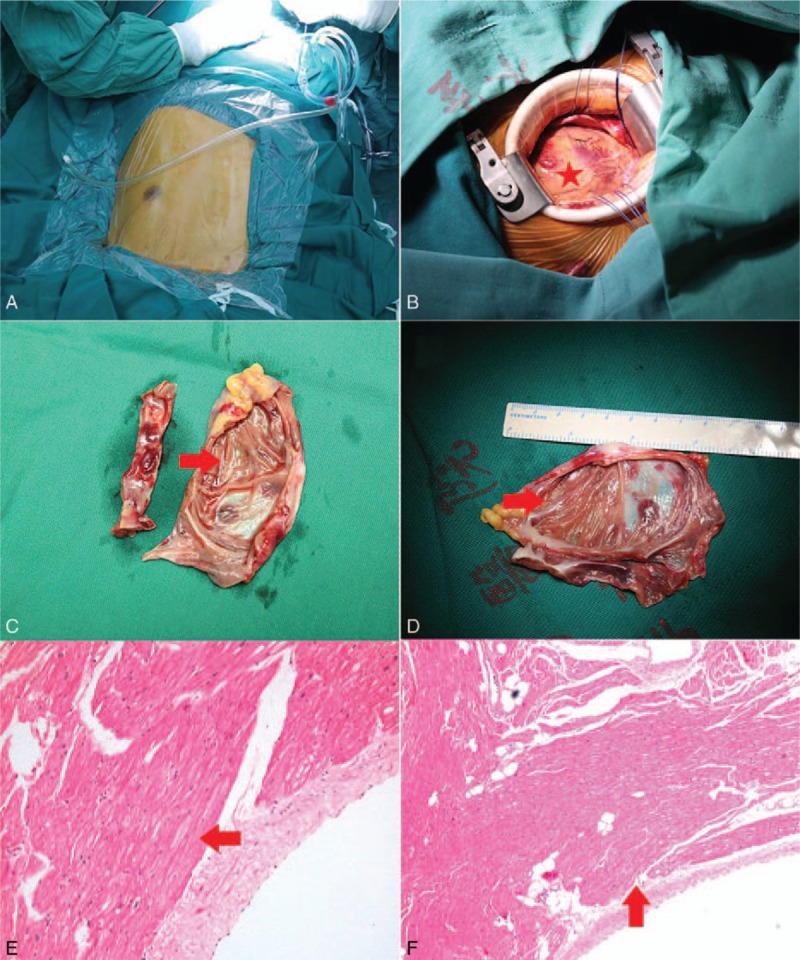
Interventions: The patient underwent an aneurysmectomy without any complications.
Outcomes: TTE confirmed the disappearance of the LAAA from the left parasternal short-axis view of the aortic root postoperatively. The patient remained asymptomatic without any adverse events at his 3-month follow-up visits.
Lessons: The associated high risk of life-threatening complications and the relative ease of surgical removal suggest that prompt evaluation should be considered in patients with lesions adjacent to the left heart border.
Copyright © 2017 The Authors. Published by Wolters Kluwer Health, Inc. All rights reserved.
Conflict of interest statement
The authors have no conflicts of interest to disclose.
Figures
References
-
- Parmley LF. Congenital atriomegaly. Circulation 1962;25:553. - PubMed
-
- Aryal MR, Hakim FA, Ghimire S, et al. Left atrial appendage aneurysm: a systematic review of 82 cases. Echocardiography 2014;31:1312–8. - PubMed
-
- Gold JP, Afifi HY, Ko W, et al. Congential giant aneurysms of the left atrial appendage: diagnosis and management. J Card Surg 1996;11:147–50. - PubMed
-
- Rikitake K, Minato N, Ohnishi H, et al. Mitral valve replacement through a giant left atrial appendage. J Cardiovasc Surg (Torino) 1999;40:127–9. - PubMed
Publication types
MeSH terms
LinkOut - more resources
Full Text Sources
Other Literature Sources
Molecular Biology Databases
