Hepatoblastoma with pure fetal epithelial differentiation in a 10-year-old boy: A rare case report and review of the literature
- PMID: 29480877
- PMCID: PMC5943836
- DOI: 10.1097/MD.0000000000009647
Hepatoblastoma with pure fetal epithelial differentiation in a 10-year-old boy: A rare case report and review of the literature
Abstract
Rationale: Hepatoblastoma is a rare malignant embryonal tumor that only accounts for approximately 1% of all pediatric cancers and mostly develops in children younger than 5 years old. Moreover, the occurrence of hepatoblastoma in adults is extremely rare.
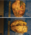
Patient concerns: Herein, we present a rare case of hepatoblastoma with pure epithelial differentiation in a 10-year-old boy.Pathological examination was performed. The tumor was 15 cm × 15 cm in size with clear margins. The cut surface was multiple nodular and grey-yellow. Histologically, the small cuboidal tumor cells were arranged in trabeculae with 2-3 cell layers. The tumor cells had eosinophilic or clear cytoplasm, formed dark and light areas, and were positive for alpha-fetoprotein, CK, CK8/18, CD10, hepatocyte, and GPC3. CD34 staining revealed that the sinusoids were lined by endothelial cells in the tumor tissues. The Ki67 index was approximately 20%.
Diagnoses: Based on these findings, the case was diagnosed as hepatoblastoma with pure fetal epithelial differentiation.
Interventions: The tumor was completely removed.
Outcomes: No recurrence was found 3 months after the operation.
Lessons: Hepatoblastoma with pure epithelial differentiation can also occur in older children. Children rarely notice and report any physical abnormality, and this may be among the primary reasons for the late diagnosis of the tumor. Annual heath checks may be beneficial in the detection of these rare tumors and improvement of patient outcomes.
Copyright © 2017 The Authors. Published by Wolters Kluwer Health, Inc. All rights reserved.
Conflict of interest statement
The authors declare that they have no competing interests.
Figures


Similar articles
-
Coexisting Infantile Hepatic Hemangioma and Hepatoblastoma in a Neonate: A Case Report.Int J Surg Pathol. 2023 Jun;31(4):485-490. doi: 10.1177/10668969231171127. Epub 2023 Apr 25. Int J Surg Pathol. 2023. PMID: 37097887
-
A rare case of adult hepatoblastoma with neuroendocrine differentiation misdiagnosed as neuroendocrine tumor.Int J Clin Exp Pathol. 2013;6(2):308-13. Epub 2013 Jan 15. Int J Clin Exp Pathol. 2013. PMID: 23330017 Free PMC article.
-
[Glypican 3 expression in hepatoblastoma and its diagnostic implication].Zhonghua Bing Li Xue Za Zhi. 2013 Dec;42(12):806-9. Zhonghua Bing Li Xue Za Zhi. 2013. PMID: 24507097 Chinese.
-
[Epithelial hepatoblastomas in the adult].Rev Gastroenterol Mex. 1994 Jul-Sep;59(3):231-5. Rev Gastroenterol Mex. 1994. PMID: 7716366 Review. Spanish.
-
Mixed hepatoblastoma in an adult--a case report and literature review.J Korean Med Sci. 1997 Aug;12(4):369-73. doi: 10.3346/jkms.1997.12.4.369. J Korean Med Sci. 1997. PMID: 9288639 Free PMC article. Review.
Cited by
-
Two Cases of Hepatoblastoma in Adults.Clin Pathol. 2022 Oct 26;15:2632010X221129592. doi: 10.1177/2632010X221129592. eCollection 2022 Jan-Dec. Clin Pathol. 2022. PMID: 36313585 Free PMC article.
-
PCSK9 in Liver Cancers at the Crossroads between Lipid Metabolism and Immunity.Cells. 2022 Dec 19;11(24):4132. doi: 10.3390/cells11244132. Cells. 2022. PMID: 36552895 Free PMC article. Review.
-
SNORA14A inhibits hepatoblastoma cell proliferation by regulating SDHB-mediated succinate metabolism.Cell Death Discov. 2023 Jan 30;9(1):36. doi: 10.1038/s41420-023-01325-0. Cell Death Discov. 2023. PMID: 36717552 Free PMC article.
-
Metastatic Hepatoblastoma in Adolescence: A Clinical Case Report and Literature Review.Cureus. 2024 Apr 17;16(4):e58460. doi: 10.7759/cureus.58460. eCollection 2024 Apr. Cureus. 2024. PMID: 38765389 Free PMC article.
References
-
- Herzog CE, Andrassy RJ, Eftekhari F. Childhood cancers: hepatoblastoma. Oncologist 2000;5:445–53. - PubMed
-
- Bosman FT, Carneiro F, Hruban RH, et al. WHO Classification of Tumors of the Digestive System. 4th ed.Lyon:IRAC; 2015.
-
- Caso-Maestro Ó, Justo-Alonso I, Cambra-Molero F, et al. Adult hepatoblastoma: case report with adrenal recurrence. Rev Esp Enferm Dig 2013;105:638–9. - PubMed
-
- Iacob D, Serban A, Fufezan O, et al. Mixed hepatoblastoma in child. Case report. Med Ultrason 2010;12:157–62. - PubMed
Publication types
MeSH terms
LinkOut - more resources
Full Text Sources
Other Literature Sources
Medical
Research Materials

