

Chronic E-Cigarette Exposure Alters the Human Bronchial Epithelial Proteome
- PMID: 29481290
- PMCID: PMC6034122
- DOI: 10.1164/rccm.201710-2033OC
Chronic E-Cigarette Exposure Alters the Human Bronchial Epithelial Proteome
Abstract
Rationale: E-cigarettes vaporize propylene glycol/vegetable glycerin (PG/VG), nicotine, and flavorings. However, the long-term health effects of exposing lungs to vaped e-liquids are unknown.
Objectives: To determine the effects of chronic vaping on pulmonary epithelia.
Methods: We performed research bronchoscopies on healthy nonsmokers, cigarette smokers, and e-cigarette users (vapers) and obtained bronchial brush biopsies and lavage samples from these subjects for proteomic investigation. We further employed in vitro and murine exposure models to support our human findings.
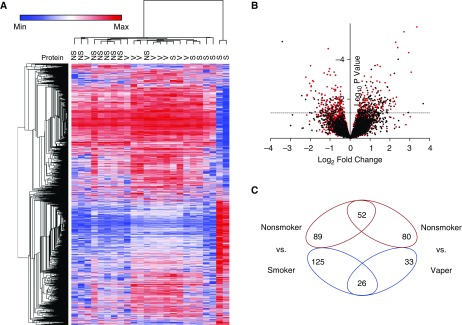
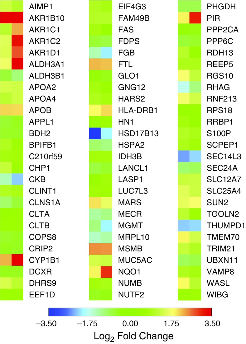
Measurements and main results: Visual inspection by bronchoscopy revealed that vaper airways appeared friable and erythematous. Epithelial cells from biopsy samples revealed approximately 300 proteins that were differentially expressed in smoker and vaper airways, with only 78 proteins being commonly altered in both groups and 113 uniquely altered in vapers. For example, CYP1B1 (cytochrome P450 family 1 subfamily B member 1), MUC5AC (mucin 5 AC), and MUC4 levels were increased in vapers. Aerosolized PG/VG alone significantly increased MUC5AC protein in human airway epithelial cultures and in murine nasal epithelia in vivo. We also found that e-liquids rapidly entered cells and that PG/VG reduced membrane fluidity and impaired protein diffusion.
Conclusions: We conclude that chronic vaping exerts marked biological effects on the lung and that these effects may in part be mediated by the PG/VG base. These changes are likely not harmless and may have clinical implications for the development of chronic lung disease. Further studies will be required to determine the full extent of vaping on the lung.
Keywords: chronic obstructive pulmonary disease; mucin; tobacco; vaping.
Figures


Comment in
-
Effects of E-Cigarette Use on Human Lung Tissue. On Harm Reduction and Causing Harm.Am J Respir Crit Care Med. 2018 Jul 1;198(1):6-7. doi: 10.1164/rccm.201802-0299ED. Am J Respir Crit Care Med. 2018. PMID: 29518342 No abstract available.
-
Reply to Shields et al.: Electronic Cigarettes and the Lung Proteome.Am J Respir Crit Care Med. 2018 Nov 15;198(10):1351-1352. doi: 10.1164/rccm.201807-1336LE. Am J Respir Crit Care Med. 2018. PMID: 30153039 Free PMC article. No abstract available.
-
Electronic Cigarettes and the Lung Proteome.Am J Respir Crit Care Med. 2018 Nov 15;198(10):1350-1351. doi: 10.1164/rccm.201806-1151LE. Am J Respir Crit Care Med. 2018. PMID: 30153045 Free PMC article. No abstract available.
References
-
- Grana R, Benowitz N, Glantz S. Background paper on e-cigarettes (electronic nicotine delivery systems) San Francisco: UCSF Center for Tobacco Control Research and Education; 2013. [accessed 2018 Jan 1]. Available from: https://escholarship.org/uc/item/13p2b72n.
-
- Benowitz NL. Pharmacology of nicotine: addiction and therapeutics. Annu Rev Pharmacol Toxicol. 1996;36:597–613. - PubMed
-
- Caramori G, Adcock IM, Casolari P, Ito K, Jazrawi E, Tsaprouni L, et al. Unbalanced oxidant-induced DNA damage and repair in COPD: a link towards lung cancer. Thorax. 2011;66:521–527. - PubMed
-
- Messner B, Bernhard D. Smoking and cardiovascular disease: mechanisms of endothelial dysfunction and early atherogenesis. Arterioscler Thromb Vasc Biol. 2014;34:509–515. - PubMed
Publication types
MeSH terms
Substances
Grants and funding
LinkOut - more resources
Full Text Sources
Other Literature Sources
Medical
