. 2018 Feb 26;13(2):e0191264.
doi: 10.1371/journal.pone.0191264. eCollection 2018.

17-AAG inhibits vemurafenib-associated MAP kinase activation and is synergistic with cellular immunotherapy in a murine melanoma model

Affiliations

17-AAG inhibits vemurafenib-associated MAP kinase activation and is synergistic with cellular immunotherapy in a murine melanoma model

Sandeep S Joshi et al. PLoS One. .

Abstract

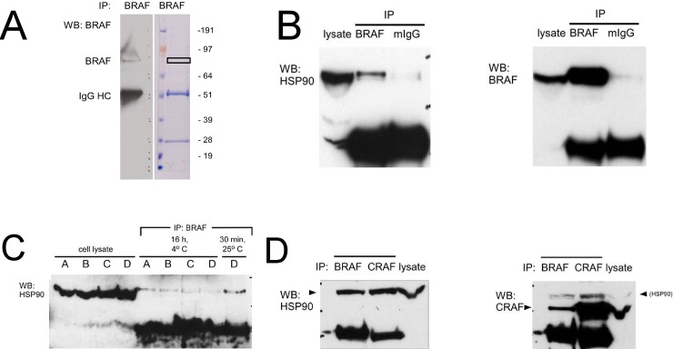
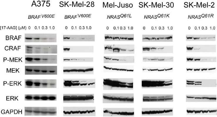
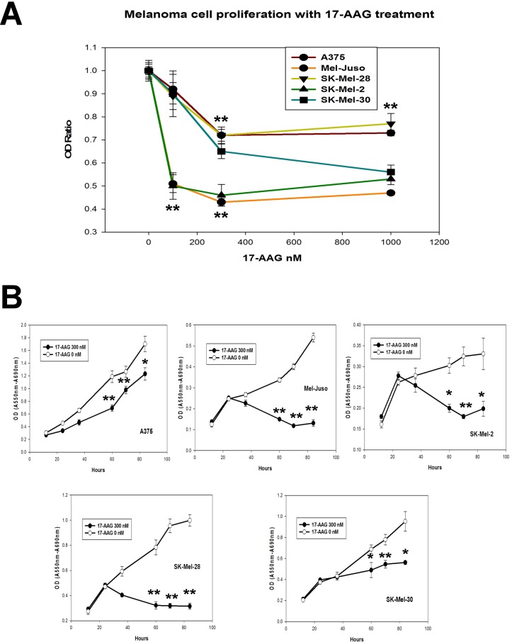
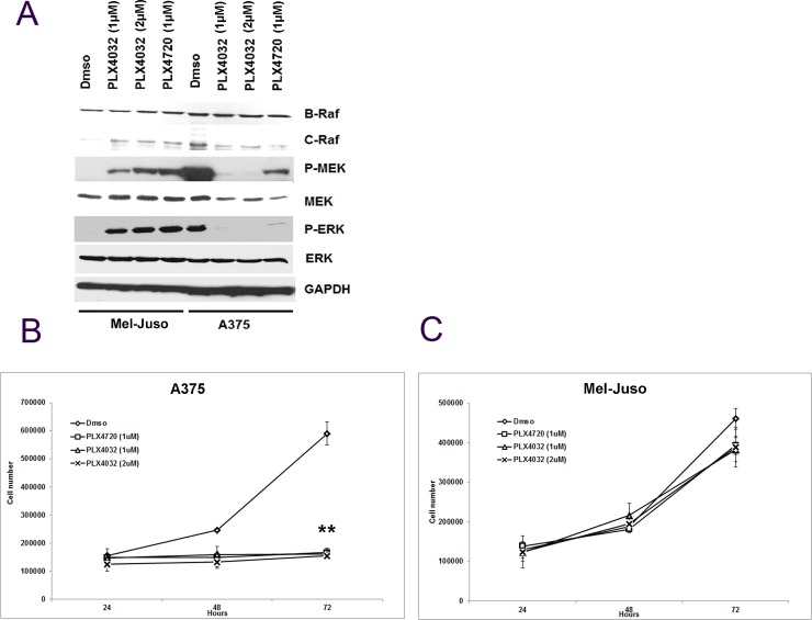
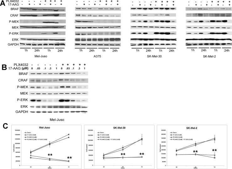
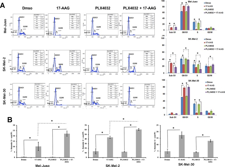
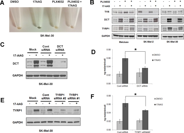
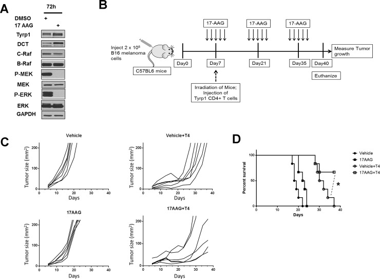
Heat shock protein 90 (HSP90) is a molecular chaperone which stabilizes client proteins with important roles in tumor growth. 17-allylamino-17-demethoxygeldanamycin (17-AAG), an inhibitor of HSP90 ATPase activity, occupies the ATP binding site of HSP90 causing a conformational change which destabilizes client proteins and directs them towards proteosomal degradation. Malignant melanomas have active RAF-MEK-ERK signaling which can occur either through an activating mutation in BRAF (BRAFV600E) or through activation of signal transduction upstream of BRAF. Prior work showed that 17-AAG inhibits cell growth in BRAFV600E and BRAF wildtype (BRAFWT) melanomas, although there were conflicting reports about the dependence of BRAFV600E and BRAFWT upon HSP90 activity for stability. Here, we demonstrate that BRAFWT and CRAF are bound by HSP90 in BRAFWT, NRAS mutant melanoma cells. HSP90 inhibition by 17-AAG inhibits ERK signaling and cell growth by destabilizing CRAF but not BRAFWT in the majority of NRAS mutant melanoma cells. The highly-selective BRAFV600E inhibitor, PLX4032 (vemurafenib), inhibits ERK signaling and cell growth in mutant BRAF melanoma cells, but paradoxically enhances signaling in cells with wild-type BRAF. In our study, we examined whether 17-AAG could inhibit PLX4032-enhanced ERK signaling in BRAFWT melanoma cells. As expected, PLX4032 alone enhanced ERK signaling in the BRAFWT melanoma cell lines Mel-Juso, SK-Mel-2, and SK-Mel-30, and inhibited signaling and cell growth in BRAFV600E A375 cells. However, HSP90 inhibition by 17-AAG inhibited PLX4032-enhanced ERK signaling and inhibited cell growth by destabilizing CRAF. Surprisingly, 17-AAG also stimulated melanin production in SK-Mel-30 cells and enhanced TYRP1 and DCT expression without stimulating TYR production in all three BRAFWT cell lines studied as well as in B16F10 mouse melanoma cells. In vivo, the combination of 17-AAG and cellular immunotherapy directed against Tyrp1 enhanced the inhibition of tumor growth compared to either therapy alone. Our studies support a role for 17-AAG and HSP90 inhibition in enhancing cellular immunotherapy for melanoma.


Conflict of interest statement

Competing Interests: The authors have declared that no competing interests exist.

Figures

Fig 1
Fig 1. Molecular interactions between BRAF, CRAF, and HSP90 in human melanoma cells.
(A) Mass spectroscopic identification of HSP90α and HSP90β as a binding partner of BRAF in human melanoma cells. An immunoprecipitate of BRAF from Mel-Juso melanoma cell lysate was incubated with an anti-BRAF monoclonal antibody, electrophoresed, and subjected to Western blot analysis (left) and Coomassie Blue staining (right). Excision of an ~85 kDa Coomassie-stained band (box) followed by mass spectroscopic analysis revealed peptides corresponding of α- and β-isoforms of HSP90. (B) Co-immunoprecipitation of HSP90 from human melanoma cell lysate with mouse monoclonal anti-BRAF. Following electrophoresis of an anti-BRAF immunoprecipitate, Western blotting (left panel) with monoclonal anti-HSP90 demonstrates HSP90 in the immunoprecipitation complex (BRAF) compared to a control immunoprecipitate with murine IgG (mIgG). (Right panel) Reprobing the membrane in (left panel) with anti-BRAF (right panel) confirms the presence of BRAF in the immunoprecipitate. (C) Immunoprecipitation conditions on stability of HSP90-BRAF interaction. Monoclonal anti-BRAF was incubated with Mel-Juso cell lysate under the following conditions: Condition A, PBS, pH 7.4, 0.1% SDS, 0.5% sodium deoxycholate; Condition B, 50 mM Tris-HCl, pH 7.4, 0.15 M NaCl, 1% NP-40, 0.25% sodium deoxycholate; Condition C, 50 mM Tris-HCl, pH 7.4, 0.1 M NaCl, 1% NP-40 (TENSV) (5); Condition D, 10 mM HEPES, pH 7.35, 20 mM sodium molybdate [31]. (D) (Left panel) Co-immunoprecipitation of HSP90 with either BRAF or CRAF in human melanoma cells. Mel-Juso cell lysate was incubated with either monoclonal anti-BRAF or anti-CRAF. Following electrophoresis and Western transfer, the blot was reprobed with anti-HSP90. (Right panel) Co-immunoprecipitation of CRAF with BRAF. The blot after stripping was reprobed with anti-CRAF. The faint band above the CRAF band is residual signal from previous probing with anti-HSP90.
Fig 2
Fig 2. Effects of 17-AAG upon BRAF and CRAF stability and on MAP kinase signaling in human melanoma cells.
Human melanoma cells (A375, SK-Mel-28, Mel-Juso, SK-Mel-30, and SK-Mel-2) were incubated with increasing concentrations of 17-AAG (0.1, 0.3, 1.0 μM) for 24 h, and the cell lysates collected were examined for phosphorylation of MAPK pathway by western blot.
Fig 3
Fig 3. Effect of 17-AAG on cell proliferation in human melanoma cells.
(A) Inhibition of melanoma cell viability with increasing concentrations of 17-AAG. Human melanoma cells (A375, SK-Mel-28, Mel-Juso, SK-Mel-30, and SK-Mel-2) were incubated with increasing concentrations (0, 0.1, 0.3, 1.0 μM) of 17-AAG for 48 h. Relative cell number was assessed by differential absorbance at 550 and 690 nm using the MTT assay. (B) Time-dependent growth inhibition of human melanoma cells with 0.3 μM 17-AAG. Human melanoma cells lines were incubated with 0.3 μM 17-AAG for 12, 24, 36, 60, 70, and 84 h before determination of relative cell number using the MTT assay. (**P<0.01; *P<0.05).
Fig 4
Fig 4. Effect of PLX4032 on MAPK signaling and on proliferation of BRAFWT and BRAFV600E human melanoma cells.
(A) Cultured human melanoma cells Mel-Juso and A375 were incubated with PLX4032 (1μM or 2μM) or PLX4720 (1μM) or vehicle (DMSO) for 24h, and cell lysates were studied for protein and phosphoprotein expression by western blot. (B) Human melanoma cell lines Mel-Juso and A375 were incubated with PLX4032 (1μM or 2μM) or PLX4720 (1μM) or vehicle (DMSO) for 24, 48, and 72h. Time-dependent inhibition of PLX4032 and PLX4720 on cell proliferation was determined by counting cells that exclude Trypan blue.
Fig 5
Fig 5. 17-AAG treatment inhibits PLX4032 enhanced MAPK signaling in BRAFWT human melanoma cells.
(A) Human melanoma cells (Mel-Juso, A375, SK-Mel-30 and SK-Mel-2) were pre-incubated with PLX4032 (2μM) or vehicle (DMSO) for 24h and cells were further incubated with17-AAG (1μM) for 1h or 24h. Cell lysates were examined for the phosphorylation pattern of MAPK pathway by western blot. (B) Cultured human melanoma cell line Mel-Juso was pre-incubated with PLX4032 (2μM) or vehicle (DMSO) for 24h and these cells were subsequently treated with increasing concentrations of 17-AAG (0, 0.03, 0.1, 0.3 or 1μM) for 24h. Cell lysates were collected and studied for phosphoprotein levels of MAPK pathway. (C) Human melanoma cell lines Mel-Juso, SK-Mel-30 and SK-Mel-2 were pre-incubated with PLX4032 (2μM) or vehicle (DMSO) for 24h and were further treated with or without 17-AAG for 24, 48, and 72h. Time-dependent inhibition of human melanoma cell growth with PLX4032 and 17-AAG was determined by counting cells that exclude Trypan blue.
Fig 6
Fig 6. Inhibitory effects of 17-AAG on cell cycle arrest and apoptosis of PLX4032 pretreated human melanoma cells.
(A) (left panel) Cell cycle distribution was determined by flow cytometry, on Mel-Juso, SK-Mel-2 and SK-Mel-30 cells pre-incubated with PLX4032 (2μM) or vehicle (DMSO) for 24h with subsequent incubation with or without 17-AAG (1μM) for an additional 72h. Cell cycle profiles were obtained by cell population-based DNA content analysis by flow cytometry (propidium iodide staining) of treated and untreated cells. (Right panel) Graphical representation of the data. The experiment in (left panel) was performed in triplicate and the percent of cells in Sub G1, G0/G1, S, and G2/M was quantified. Data (mean ± SD, n = 3); *P<0.01. (B) Effect of apoptosis was determined by flow cytometry on human melanoma cells (Mel-Juso, SK-Mel-2, SK-Mel-30) pre-incubated with PLX4032 (2μM) or vehicle (DMSO) for 24h, with subsequent incubation with or without 17-AAG (1μM) for an additional 72h. The data is a mean of percentage of Annexin V-positive and PI-negative cell population from three independent experiments. *P<0.01.
Fig 7
Fig 7. HSP90 inhibition by 17AAG induces pigmentation in human melanoma cells.
(A) SK-Mel-30 human melanoma cells pretreated with or without PLX4032 were further treated with 17-AAG or vehicle for 72h, and then harvested and spun down. Data shows the cell pellet color.17-AAG alone induced pigmentation in human melanoma cells. (B) Human melanoma cells (Mel-Juso, A375, SK-Mel-30 and SK-Mel-2) were pre-incubated with PLX4032 (2μM) or vehicle (DMSO) for 24h and subsequently incubated with or without 17-AAG (1μM) for 48h. Cell lysates were collected and studied for the expression of TYR, DCT and TYRP1 proteins by western blot. Data show 17AAG induced DCT and TYRP1 protein expression regardless of PLX4032 treatments whereas TYR was either unchanged (Mel-Juso, SK-Mel-30) or decreased (SK-Mel-2). (C) SK-mel-30 melanoma cells transfected with a control siRNA or DCT siRNA for 24h were incubated with or without 17-AAG for 48h. DCT knockdown was confirmed by western blot. (D) SK-mel-30 melanoma cells transfected with DCT siRNA show significant reduction in melanin content in 17AAG-treated cells in comparison to control siRNA treated cells. Data are represented as percentage of control and SD, measured from three independent experiments. *P < 0.05 vs. control siRNA + 17-AAG. (E) SK-Mel-30 melanoma cells transfected with a control siRNA or 2 different TYRP1 siRNAs (#1, #2) for 24h. They were further treated with or without 17-AAG for 48h. Knockdown of TYRP1 expression was confirmed by collecting cell lysates and running a western blot. (F) SK-Mel-30 melanoma cells transfected with TYRP1 siRNA #2 shows significant reduction in melanin content in 17AAG treated cells in comparison to control siRNA. Data are represented as percentage of control and SD, measured from three independent experiments. *P < 0.05 vs. control siRNA + 17-AAG.
Fig 8
Fig 8. 17AAG promotes the inhibition of Tyrp1 specific CD4+ T cell treated melanoma tumor growth.
(A) Cultured B16 mouse melanoma cell lines were incubated with 17-AAG (1.0 μM) for 72 h, and cell lysates collected and studied for protein and phosphoprotein expression following SDS-PAGE and Western transfer. (B) The C57BL6 mice were subcutaneously injected with 2 x 10⁵ B16 mouse melanoma cells. Five days after B16 tumor cell injection, the mice received intraperitoneal injection of either 17AAG or vehicle at a dose of 75 mg/Kg body weight x 5 consecutive days. On seventh day after tumor challenge, all mice were irradiated one time with 550 rads (5.5Gy). 50% of 17AAG and 50% of Vehicle treated tumor bearing mice received 2 x 10⁵ numbers of TYRP1-specific CD4+ T cells (T4 cells) were injected intravenously. The 17-AAG dose of 75 mg/kg x 5 consecutive days was repeated every 2 weeks following initial administration of 17-AAG until the end of experiment. (C) The time dependent effect on the B16 melanoma tumor growth in C57BL6 mice treated either with Vehicle, 17AAG, Vehicle+T4 cells or 17AAG+T4 cells were determined by measuring their growth at regular intervals till the end of the experiment (n = 6). Data is a representative of two independent experiments. (D) The survival curve analysis shows that mice receiving no T4 cell transfer and only T4 cell transfer had no surviving mice at day 40. (*P<0.05 is a comparison between Vehicle+T4 cells and 17AAG+T4 cells as indicated by dotted lines).

References

    1. Neckers L. (2007) Heat shock protein 90: The cancer chaperone. J Biosci 32(3): 517–530. - PubMed
    1. Xu W, Mimnaugh E, Rosser MF, Nicchitta C, Marcu M, et al. (2001) Sensitivity of mature Erbb2 to geldanamycin is conferred by its kinase domain and is mediated by the chaperone protein Hsp90. J Biol Chem 276(5): 3702–3708. doi: 10.1074/jbc.M006864200 - DOI - PubMed
    1. Basso AD, Solit DB, Chiosis G, Giri B, Tsichlis P, et al. (2002) Akt forms an intracellular complex with heat shock protein 90 (Hsp90) and Cdc37 and is destabilized by inhibitors of Hsp90 function. J Biol Chem 277(42): 39858–39866. doi: 10.1074/jbc.M206322200 - DOI - PubMed
    1. Schulte TW, Blagosklonny MV, Ingui C, Neckers L. (1995) Disruption of the raf-1-Hsp90 molecular complex results in destabilization of raf-1 and loss of raf-1-ras association. J Biol Chem 270(41): 24585–24588. - PubMed
    1. Schulte TW, Blagosklonny MV, Romanova L, Mushinski JF, Monia BP, et al. (1996) Destabilization of raf-1 by geldanamycin leads to disruption of the raf-1-MEK-mitogen-activated protein kinase signalling pathway. Mol Cell Biol 16(10): 5839–5845. - PMC - PubMed

Publication types

MeSH terms