

Scene and human face recognition in the central vision of patients with glaucoma
- PMID: 29481572
- PMCID: PMC5826536
- DOI: 10.1371/journal.pone.0193465
Scene and human face recognition in the central vision of patients with glaucoma
Abstract
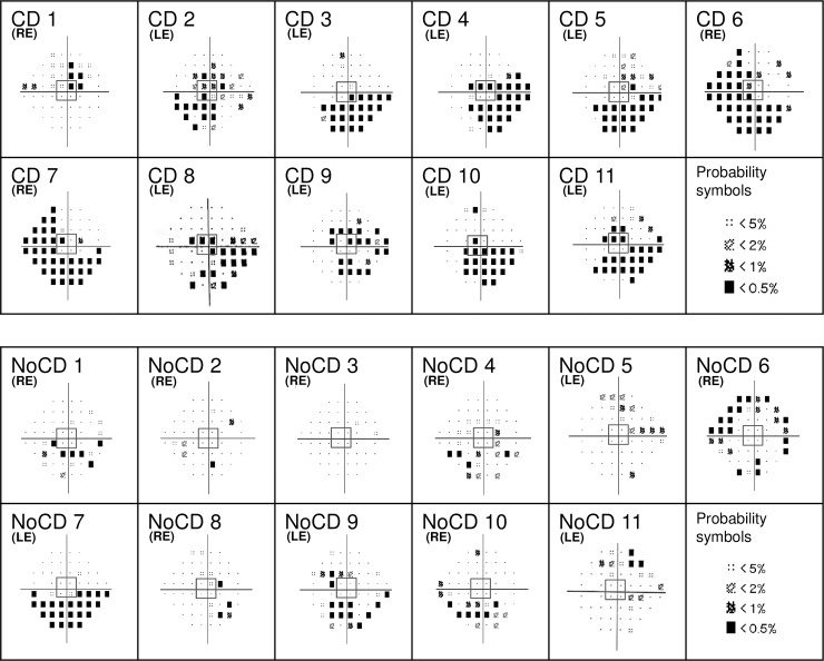
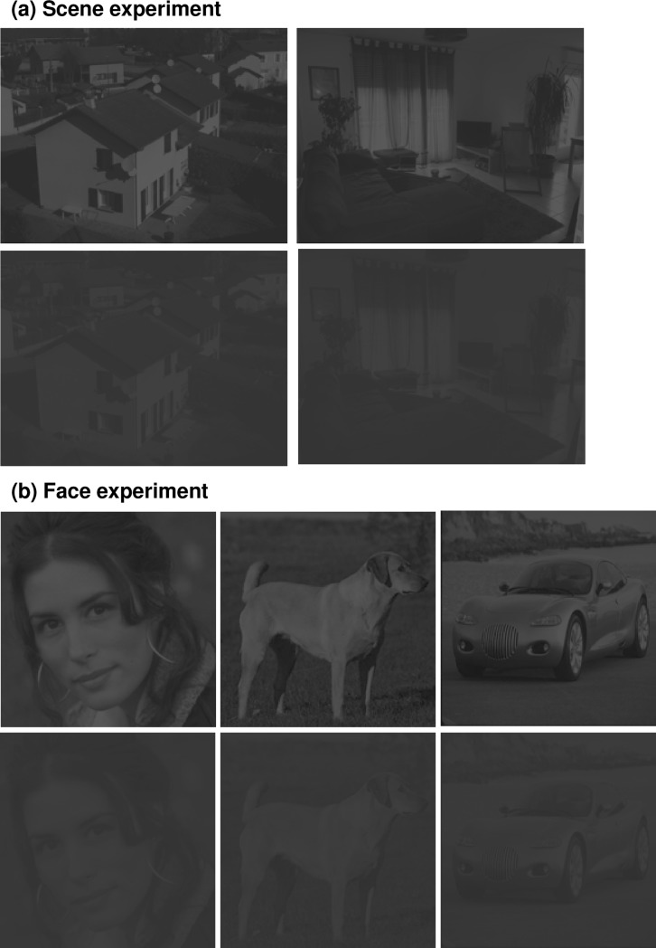
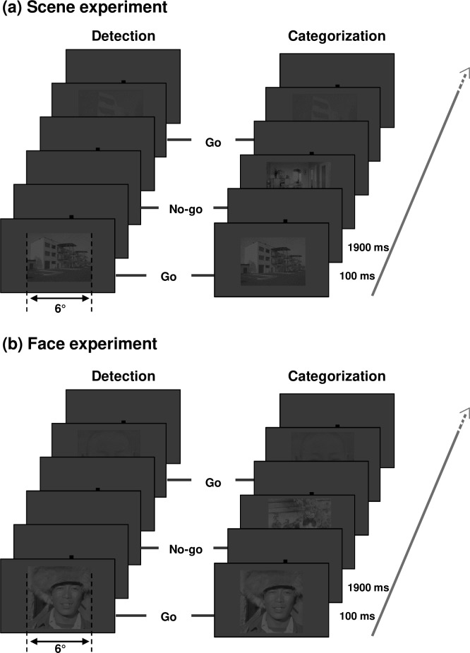
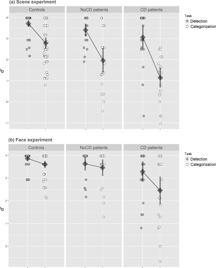
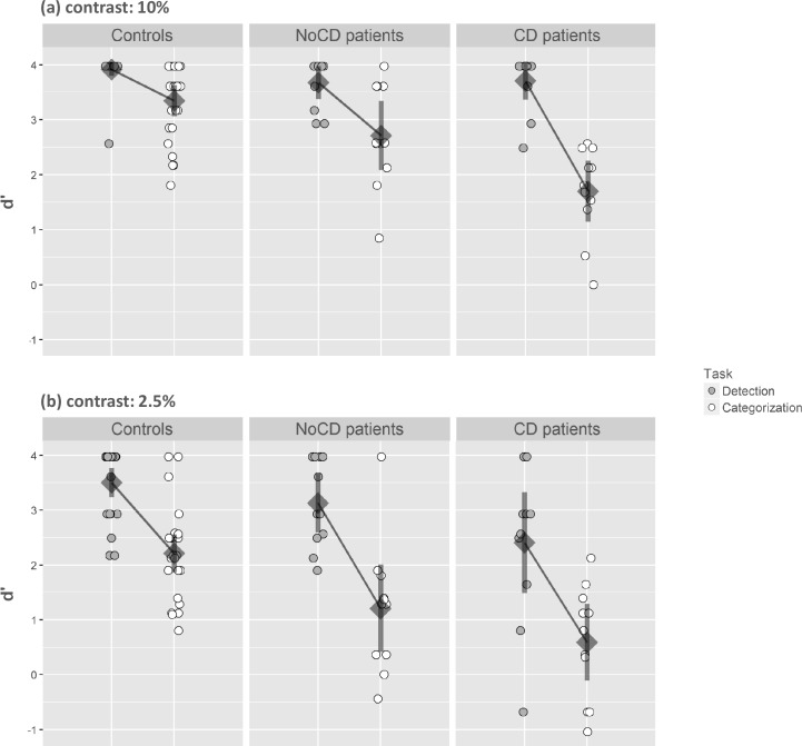
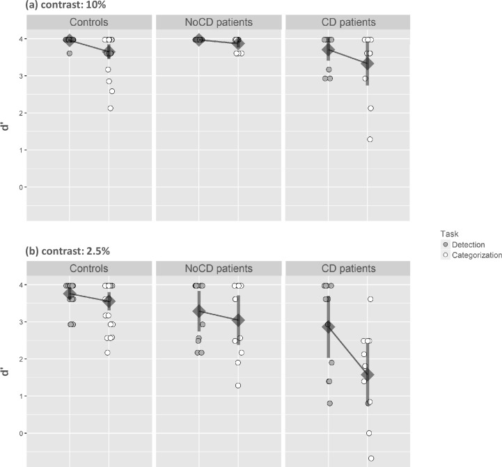
Primary open-angle glaucoma (POAG) firstly mainly affects peripheral vision. Current behavioral studies support the idea that visual defects of patients with POAG extend into parts of the central visual field classified as normal by static automated perimetry analysis. This is particularly true for visual tasks involving processes of a higher level than mere detection. The purpose of this study was to assess visual abilities of POAG patients in central vision. Patients were assigned to two groups following a visual field examination (Humphrey 24-2 SITA-Standard test). Patients with both peripheral and central defects and patients with peripheral but no central defect, as well as age-matched controls, participated in the experiment. All participants had to perform two visual tasks where low-contrast stimuli were presented in the central 6° of the visual field. A categorization task of scene images and human face images assessed high-level visual recognition abilities. In contrast, a detection task using the same stimuli assessed low-level visual function. The difference in performance between detection and categorization revealed the cost of high-level visual processing. Compared to controls, patients with a central visual defect showed a deficit in both detection and categorization of all low-contrast images. This is consistent with the abnormal retinal sensitivity as assessed by perimetry. However, the deficit was greater for categorization than detection. Patients without a central defect showed similar performances to the controls concerning the detection and categorization of faces. However, while the detection of scene images was well-maintained, these patients showed a deficit in their categorization. This suggests that the simple loss of peripheral vision could be detrimental to scene recognition, even when the information is displayed in central vision. This study revealed subtle defects in the central visual field of POAG patients that cannot be predicted by static automated perimetry assessment using Humphrey 24-2 SITA-Standard test.
Conflict of interest statement
Figures
Similar articles
-
Humphrey matrix frequency doubling perimetry for detection of visual-field defects in open-angle glaucoma.Br J Ophthalmol. 2009 May;93(5):582-8. doi: 10.1136/bjo.2007.119909. Epub 2008 Jul 31. Br J Ophthalmol. 2009. PMID: 18669543
-
Diffuse and localised visual field defects to automated perimetry in primary open angle glaucoma.Eye (Lond). 1995;9 ( Pt 6):745-50. doi: 10.1038/eye.1995.188. Eye (Lond). 1995. PMID: 8849543
-
The contribution of central and peripheral vision in scene categorization: a study on people with central vision loss.Vision Res. 2014 May;98:46-53. doi: 10.1016/j.visres.2014.03.004. Epub 2014 Mar 20. Vision Res. 2014. PMID: 24657253
-
[Aiming for zero blindness].Nippon Ganka Gakkai Zasshi. 2015 Mar;119(3):168-93; discussion 194. Nippon Ganka Gakkai Zasshi. 2015. PMID: 25854109 Review. Japanese.
-
Peripheral visual field loss and activities of daily living.Curr Opin Neurol. 2023 Feb 1;36(1):19-25. doi: 10.1097/WCO.0000000000001125. Epub 2022 Nov 21. Curr Opin Neurol. 2023. PMID: 36409221 Review.
Cited by
-
MNREAD Reading Vision in Adults With Glaucoma Under Mesopic and Photopic Conditions.Invest Ophthalmol Vis Sci. 2023 Dec 1;64(15):43. doi: 10.1167/iovs.64.15.43. Invest Ophthalmol Vis Sci. 2023. PMID: 38153749 Free PMC article.
-
Reduced functional connectivity between central representations of V1 and foveal-biased face-selective region in central vision loss.Brain Struct Funct. 2025 Jul 3;230(6):111. doi: 10.1007/s00429-025-02973-x. Brain Struct Funct. 2025. PMID: 40608114 Free PMC article.
-
Impact of Glaucomatous Ganglion Cell Damage on Central Visual Function.Annu Rev Vis Sci. 2024 Sep;10(1):425-453. doi: 10.1146/annurev-vision-110223-123044. Annu Rev Vis Sci. 2024. PMID: 39292555 Free PMC article. Review.
-
Impact of glaucoma on the spatial frequency processing of scenes in central vision.Vis Neurosci. 2023 Feb 8;40:E001. doi: 10.1017/S0952523822000086. Vis Neurosci. 2023. PMID: 36752177 Free PMC article.
-
Spatial correlation between localized decreases in exploratory visual search performance and areas of glaucomatous visual field loss.Graefes Arch Clin Exp Ophthalmol. 2019 Jan;257(1):153-160. doi: 10.1007/s00417-018-4164-9. Epub 2018 Oct 27. Graefes Arch Clin Exp Ophthalmol. 2019. PMID: 30368564
References
-
- Hood DC, Raza AS, de Moraes CG V, Liebmann JM, Ritch R. Glaucomatous damage of the macula. Prog Retin Eye Res. 2013. doi: 10.1016/j.preteyeres.2012.08.003 - DOI - PMC - PubMed
-
- Crabb DP, Smith ND, Glen FC, Burton R, Garway-Heath DF. How does glaucoma look?: Patient perception of visual field loss. Ophthalmology [Internet]. 2013;120(6):1120–6. Available from: http://dx.doi.org/10.1016/j.ophtha.2012.11.043 - DOI - PubMed
-
- Crabb DP. A view on glaucoma—Are we seeing it clearly? Eye. 2016;30(2):304–13. doi: 10.1038/eye.2015.244 - DOI - PMC - PubMed
-
- Ramulu P. Glaucoma and disability: which tasks are affected, and at what stage of disease? Curr Opin Ophthalmol. 2009;20(2):92–98. doi: 10.1097/ICU.0b013e32832401a9 - DOI - PMC - PubMed
-
- McKendrick AM, Badcock DR, Morgan WH. The detection of both global motion and global form is disrupted in glaucoma. Investig Ophthalmol Vis Sci. 2005. doi: 10.1167/iovs.04-1406 - DOI - PubMed
Publication types
MeSH terms
LinkOut - more resources
Full Text Sources
Other Literature Sources
Medical
