. 2018 May 18;46(9):4699-4714.
doi: 10.1093/nar/gky116.

Turnover of aberrant pre-40S pre-ribosomal particles is initiated by a novel endonucleolytic decay pathway

Affiliations

Turnover of aberrant pre-40S pre-ribosomal particles is initiated by a novel endonucleolytic decay pathway

Elodie Choque et al. Nucleic Acids Res. .

Abstract

Ribosome biogenesis requires more than 200 trans-acting factors to achieve the correct production of the two mature ribosomal subunits. Here, we have identified Efg1 as a novel, nucleolar ribosome biogenesis factor in Saccharomyces cerevisiae that is directly linked to the surveillance of pre-40S particles. Depletion of Efg1 impairs early pre-rRNA processing, leading to a strong decrease in 18S rRNA and 40S subunit levels and an accumulation of the aberrant 23S rRNA. Using Efg1 as bait, we revealed a novel degradation pathway of the 23S rRNA. Co-immunoprecipitation experiments showed that Efg1 is a component of 90S pre-ribosomes, as it is associated with the 35S pre-rRNA and U3 snoRNA, but has stronger affinity for 23S pre-rRNA and its novel degradation intermediate 11S rRNA. 23S is cleaved at a new site, Q1, within the 18S sequence by the endonuclease Utp24, generating 11S and 17S' rRNA. Both of these cleavage products are targeted for degradation by the TRAMP/exosome complexes. Therefore, the Q1 site defines a novel endonucleolytic cleavage site of ribosomal RNA exclusively dedicated to surveillance of pre-ribosomal particles.


Figures
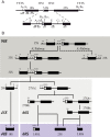
Figure 1.
Figure 1.
Simplified scheme of the pre-rRNA processing pathway in Saccharomyces cerevisiae. (A) Initial 35S pre-rRNA precursor with detailed cleavage sites as well as probes used for rRNA detection in northern blot analysis. (B) Endonucleolytic and exonucleolytic cleavages leading to the production of mature 18S, 5.8S and 25S rRNAs. Inhibition of A0, A1 and A2 cleavages (A2 pathway) leads to the non-productive A3 pathway. Resulting 23S pre-rRNA containing particles accumulate and/or are degraded. Dark and light gray boxes schematically represent 90S and 43S/66S pre-ribosomes, respectively. Mature ribosomal subunits are represented in violet.
Figure 2.
Figure 2.
Efg1 is a nucleolar protein. (A) Conserved domain in Efg1. Amino acid alignment of the DUF2361 domain of Efg1 with yeast homologs, plant and mammalian orthologs. Sequence alignments were generated using Clustal W. S. cerevisiae: Saccharomyces cerevisiae; C. glabrata: Candida glabrata; Agossypii: Ashbya gossypii; C. albicans: Candida albicans; N. crassa: Neurospora crassa; C. globossum: Candida globossum; A. thaliana: Arabidopsis thaliana; O. sativa: Oryza sativa. The putative coiled-coil region is highlighted. (B) Subcellular localization of Efg1. Yeast strain expressing Efg1-YFP and mCherry-Nop1 was grown exponentially and cell samples were used for fluorescence microscopy analysis.
Figure 3.
Figure 3.
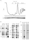
Efg1 is associated with pre-ribosomes. (A) Sedimentation profile of Efg1 on a sucrose gradient. A total extract prepared from EFG1::TAP cells growing exponentially was sedimented through a sucrose gradient, and 17 fractions were collected. The corresponding A260 profile is displayed with the characteristic annotated peaks. Each fraction was TCA precipitated, and Efg1-TAP was detected by western blotting using PAP antibodies. (B) The 35S and 23S pre-rRNAs are co-immunoprecipitated with Efg1. Northern blot analysis of (pre-)rRNAs co-precipitated with TAP-tagged Efg1 (lanes 3–4 and 7–8) or from control experiments using extracts of cells lacking a tagged protein (lanes 1–2 and 5–6). Immunoprecipitation was performed under native conditions using IgG-Sepharose. RNAs were extracted from the pellet after precipitation (lanes IP) or from total cell extract (lanes Tot) corresponding to 5% of the input used for the immunoprecipitation reactions. Following denaturing electrophoresis, RNAs were transferred to a nylon membrane and hybridized with anti-sense oligonucleotides corresponding to various (pre-)rRNAs and snoRNAs. (C) Full northern blot showing RNAs co-immunoprecipitated with Efg1 and detected using a probe hybridizing between A0 and A1 sites (1699, lanes 1 and 2) and between D and A2 (004, lanes 3 and 4).
Figure 4.
Figure 4.
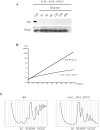
Efg1 depletion affects 40S ribosomal subunit accumulation in yeast cells. (A) Western blot analysis of 3HA-Efg1 depletion. Total proteins were extracted at the times indicated and analyzed by western blot. 3HA-Efg1 and Nhp2 were detected using anti-HA and Nhp2-specific antibodies respectively. (B) Growth rate of WT and Gal::3HA::EFG1 strains following a transfer from permissive galactose medium to glucose medium for the times indicated. Cells were maintained in exponential growth throughout the time course by dilution into pre-warmed medium. (C) Ribosome profiles of Efg1-depleted cells. GAL::3HA::EFG1 and WT BY4741 strains were grown up to 0.6 (OD600) in galactose medium and shifted to glucose for 3 h. Total cell extracts were prepared, centrifuged through 4.5–45% sucrose gradients and 17 fractions were collected. The A260 absorbance profile is presented with annotated peaks.
Figure 5.
Figure 5.
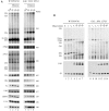
Efg1 depletion leads to a defect in A0, A1 and A2 cleavages. (A) WT BY4741 and GAL::3HA::EFG1 strains were shifted from a galactose to a glucose medium. Samples were collected before and at different times after the nutritional shift. Total RNAs were extracted from these cell samples, and the accumulation of the different pre-rRNAs, rRNAs and sn(o)RNAs was analyzed by northern blot using different probes. (B) Pulse-chase labeling of RNAs. WT BY4741 and GAL::3HA::EFG1 cells were grown in galactose containing medium and were next shifted to glucose for 3 h. Cells were then pulse labeled with [2,8–3H] adenine for 2 min. Samples were collected 0, 1, 2, 5, 10, 20 and 30 min after addition of an excess of cold adenine. Total RNAs were extracted from these samples, separated by gel electrophoresis and transferred to a nylon membrane.
Figure 6.
Figure 6.
23S pre-rRNA is targeted by endo- and exo-nucleolytic pathways for its degradation. Pre-rRNA accumulation was monitored in (A) presence of endogenous Efg1 and compared with cells depleted for Efg1 (B). Yeast cells were grown to mid-log phase in galactose containing media. Glucose was next added within the media and cells were grown in these conditions for 3 h. Aliquots were collected and total RNAs were extracted and subjected to poly(A)+ affinity purification on oligo-dT coated beads. All RNA samples were separated on 1.2% agarose gels, transferred to a nylon membrane and hybridized with specific oligonucleotide probes. Upper panels and mid panels were hybridized with ETS1 (1699) and ITS1 (004) probes, respectively. Lower panels were hybridized with an oligonucleotide specific for PGK1 mRNA (403).
Figure 7.
Figure 7.
23S pre-rRNA is cleaved at Q1 site and the PIN domain of Utp24 is required for efficient cleavage. (A) RNAs extracted from WT or rrp6Δ strains were analyzed by primer extension using probe 1510 (schematized by the arrow). A PCR fragment containing 18S sequence was used to generate a sequencing ladder leading to the identification of Q1 site. (B) Predicted secondary structure of the 18S rRNA central domain in Saccharomyces cerevisiae. Utp24 crosslinking sites are marked on the sequence and shades indicate peak height with the highest peak shown in dark grey. Q1 site (18S 618/619) is indicated. Oligonucleotide used to map Q1 site is annotated (red arrow). (C) Northern analysis of pre-rRNA processing in the pTET::utp24–3HA strain transformed with a plasmid expressing either WT Utp24 or the PIN mutant (D68N) his-tagged Utp24 protein, or an empty vector. RNAs were isolated from mid log phase cells grown 8 h in presence of doxycycline to deplete endogenous Utp24 protein. Aliquots were collected and total RNAs were extracted and subjected to poly(A)+ affinity purification on oligo-dT-coated beads. Total RNAs (lanes 1–3 and 7–9) and poly(A)+ RNAs (lanes 4–6 and 10–12) were separated on an 1.2% agarose gel and detected by northern hybridization with specific oligonucleotide probes. Left panels (lanes 1–6) were hybridized with ITS1 probe (004), right panels (lanes 7–12) were hybridized with ETS1 probe (1699). Loading was assessed by hybridizing the PGK1 mRNA (lower panel) with probe 403.
Figure 8.
Figure 8.
Proposed model for the function of Utp24 in the ‘productive’ A2 pathway and the ‘non productive’ A3 pathway. In normal growing conditions, Utp24 cleaves at A1 and A2 sites leading to the production of the 20S pre-rRNA and subsequently the production of the mature 18S rRNA. In case of defective assembly and/or processing of early pre-ribosomal particles or if growing conditions are not optimal, A1 and A2 cleavage sites are not processed by Utp24. 35S pre-rRNA is directly cleaved at A3 site by RNase MRP. Resulting 23S pre-rRNA is targeted by Utp24 at Q1 site generating 11S and 17S'. All of them are degraded by the TRAMP/exosome pathways.

Similar articles

Cited by

References

    1. Woolford J.L. Jr., Baserga S.J.. Ribosome biogenesis in the yeast Saccharomyces cerevisiae. Genetics. 2013; 195:643–681. - PMC - PubMed
    1. Henras A.K., Plisson-Chastang C., O’Donohue M.F., Chakraborty A., Gleizes P.E.. An overview of pre-ribosomal RNA processing in eukaryotes. Enzymes. 2014; 41:169–213. - PMC - PubMed
    1. Zhang J., Harnpicharnchai P., Jakovljevic J., Tang L., Guo Y., Oeffinger M., Rout M.P., Hiley S.L., Hughes T., Woolford J.L. Jr.. Assembly factors Rpf2 and Rrs1 recruit 5S rRNA and ribosomal proteins rpL5 and rpL11 into nascent ribosomes. Genes Dev. 2007; 21:2580–2592. - PMC - PubMed
    1. Dragon F., Gallagher J.E., Compagnone-Post P.A., Mitchell B.M., Porwancher K.A., Wehner K.A., Wormsley S., Settlage R.E., Shabanowitz J., Osheim Y. et al. . A large nucleolar U3 ribonucleoprotein required for 18S ribosomal RNA biogenesis. Nature. 2002; 417:967–970. - PMC - PubMed
    1. Grandi P., Rybin V., Bassler J., Petfalski E., Strauss D., Marzioch M., Schafer T., Kuster B., Tschochner H., Tollervey D. et al. . 90S pre-ribosomes include the 35S pre-rRNA, the U3 snoRNP, and 40S subunit processing factors but predominantly lack 60S synthesis factors. Mol. Cell. 2002; 10:105–115. - PubMed

Publication types

MeSH terms