

Activation and desensitization of ionotropic glutamate receptors by selectively triggering pre-existing motions
- PMID: 29481851
- PMCID: PMC6107436
- DOI: 10.1016/j.neulet.2018.02.050
Activation and desensitization of ionotropic glutamate receptors by selectively triggering pre-existing motions
Abstract
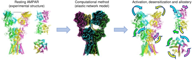
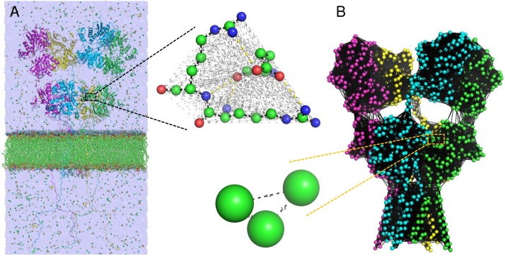
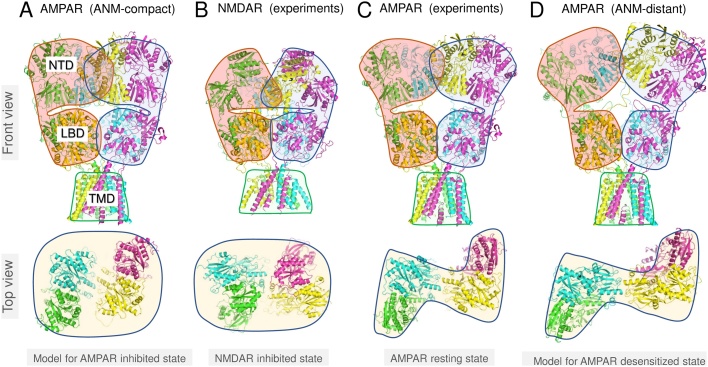
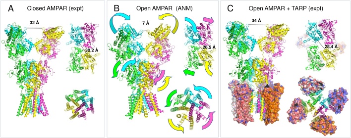
Ionotropic glutamate receptors (iGluRs) are ligand-gated ion channels that are key players in synaptic transmission and plasticity. They are composed of four subunits, each containing four functional domains, the quaternary packing and collective structural dynamics of which are important determinants of their molecular mechanism of function. With the explosion of structural studies on different members of the family, including the structures of activated open channels, the mechanisms of action of these central signaling machines are now being elucidated. We review the current state of computational studies on two major members of the family, AMPA and NMDA receptors, with focus on molecular simulations and elastic network model analyses that have provided insights into the coupled movements of extracellular and transmembrane domains. We describe the newly emerging mechanisms of activation, allosteric signaling and desensitization, as mainly a selective triggering of pre-existing soft motions, as deduced from computational models and analyses that leverage structural data on intact AMPA and NMDA receptors in different states.
Keywords: AMPA and NMDA receptors; Activation mechanism; Allosteric interactions; Desensitization; Elastic network models; Ionotropic glutamate receptors; Molecular dynamics; Simulations.
Copyright © 2018 The Authors. Published by Elsevier B.V. All rights reserved.
Figures
Similar articles
-
Cooperative Dynamics of Intact AMPA and NMDA Glutamate Receptors: Similarities and Subfamily-Specific Differences.Structure. 2015 Sep 1;23(9):1692-1704. doi: 10.1016/j.str.2015.07.002. Epub 2015 Aug 6. Structure. 2015. PMID: 26256538 Free PMC article.
-
Emerging structural insights into the function of ionotropic glutamate receptors.Trends Biochem Sci. 2015 Jun;40(6):328-37. doi: 10.1016/j.tibs.2015.04.002. Epub 2015 May 1. Trends Biochem Sci. 2015. PMID: 25941168 Free PMC article. Review.
-
Structure, Dynamics, and Allosteric Potential of Ionotropic Glutamate Receptor N-Terminal Domains.Biophys J. 2015 Sep 15;109(6):1136-48. doi: 10.1016/j.bpj.2015.06.061. Epub 2015 Aug 6. Biophys J. 2015. PMID: 26255587 Free PMC article. Review.
-
Comparative dynamics of NMDA- and AMPA-glutamate receptor N-terminal domains.Structure. 2012 Nov 7;20(11):1838-49. doi: 10.1016/j.str.2012.08.012. Epub 2012 Sep 6. Structure. 2012. PMID: 22959625 Free PMC article.
-
Functional insights from glutamate receptor ion channel structures.Annu Rev Physiol. 2013;75:313-37. doi: 10.1146/annurev-physiol-030212-183711. Epub 2012 Sep 4. Annu Rev Physiol. 2013. PMID: 22974439 Free PMC article. Review.
Cited by
-
Perception of Damaged Self in Plants.Plant Physiol. 2020 Apr;182(4):1545-1565. doi: 10.1104/pp.19.01242. Epub 2020 Jan 6. Plant Physiol. 2020. PMID: 31907298 Free PMC article. Review.
-
The structural arrangement and dynamics of the heteromeric GluK2/GluK5 kainate receptor as determined by smFRET.Biochim Biophys Acta Biomembr. 2020 Jan 1;1862(1):183001. doi: 10.1016/j.bbamem.2019.05.023. Epub 2019 Jun 11. Biochim Biophys Acta Biomembr. 2020. PMID: 31194959 Free PMC article.
-
Intrinsic dynamics is evolutionarily optimized to enable allosteric behavior.Curr Opin Struct Biol. 2020 Jun;62:14-21. doi: 10.1016/j.sbi.2019.11.002. Epub 2019 Nov 27. Curr Opin Struct Biol. 2020. PMID: 31785465 Free PMC article. Review.
-
Gating and modulation of a hetero-octameric AMPA glutamate receptor.Nature. 2021 Jun;594(7863):454-458. doi: 10.1038/s41586-021-03613-0. Epub 2021 Jun 2. Nature. 2021. PMID: 34079129 Free PMC article.
-
Computer Simulations Predict High Structural Heterogeneity of Functional State of NMDA Receptors.Biophys J. 2018 Sep 4;115(5):841-852. doi: 10.1016/j.bpj.2018.06.023. Epub 2018 Jun 28. Biophys J. 2018. PMID: 30029773 Free PMC article.
References
Publication types
MeSH terms
Substances
Grants and funding
LinkOut - more resources
Full Text Sources
Other Literature Sources
