. 2018 Mar 1;30(1):34-41.
doi: 10.5792/ksrr.17.035.

Clinical and Radiological Results with Second-Look Arthroscopic Findings after Open Wedge High Tibial Osteotomy without Arthroscopic Procedures for Medial Meniscal Root Tears

Affiliations

Clinical and Radiological Results with Second-Look Arthroscopic Findings after Open Wedge High Tibial Osteotomy without Arthroscopic Procedures for Medial Meniscal Root Tears

Hyun Il Lee et al. Knee Surg Relat Res. .

Abstract

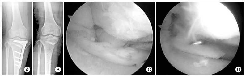
Purpose: To identify the structural integrity of the healing site after medial open wedge high tibial osteotomy (MOWHTO) in patients with a posterior root tear of the medial meniscus (PRTMM) and chondral lesion by second-look arthroscopy and to determine the clinical and radiological findings.

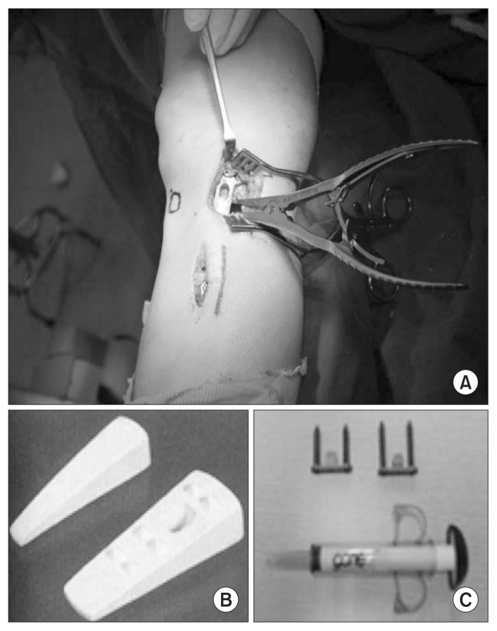
Materials and methods: From August 2010 to June 2016, 52 consecutive patients underwent MOWHTO and arthroscopic examination without a chondral resurfacing procedure and meniscal treatment for PRTMM. Twenty-four patients were available for second-look arthroscopic evaluation. The mean follow-up period was 19.5 months (range, 5 to 46 months). Clinical evaluation was based on the Lysholm knee scores and Hospital for Special Surgery (HSS) scores.

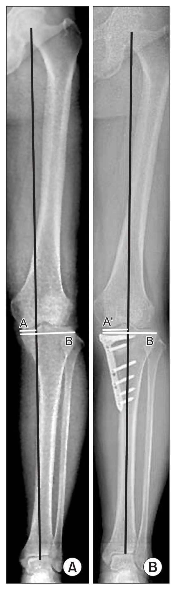
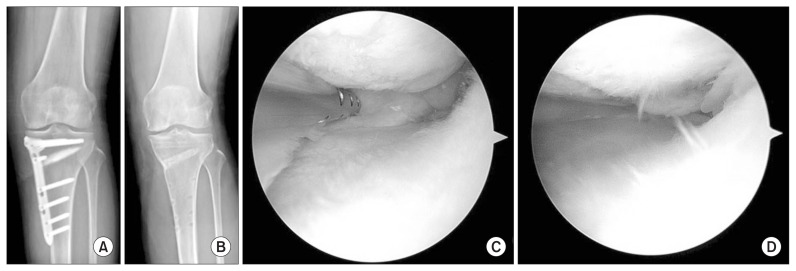
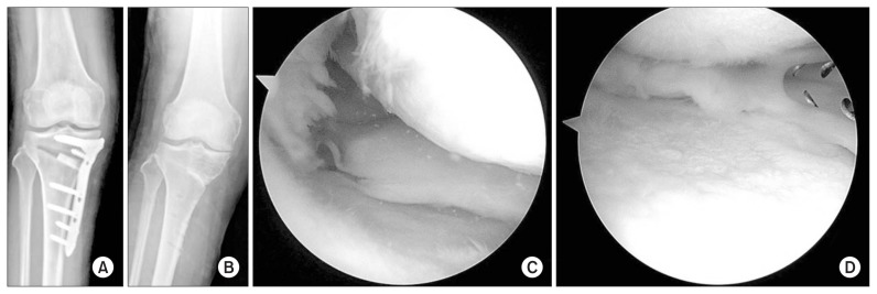
Results: There were 5 lax healing, 6 scar tissue, 13 failed healing of PRTMM. Definite change of chondral lesion was not observed. The Kellgren-Lawrence grade did not improve according to the follow-up plain radiograph. The mean Lysholm score improved from 34.7 preoperatively to 77.1 at the last follow-up, and the mean HSS score significantly increased from 36.5 to 82.4.

Conclusions: This study revealed a low rate of healing potency of PRTMM and chondral lesion after MOWHTO without any attempt for meniscal treatment or chondral resurfacing. The cartilage and healing status of PRTMM was not associated with improved clinical outcomes and radiological findings.

Keywords: Knee; Medial meniscus; Open wedge; Osteoarthritis; Osteotomy; Root tear.


Conflict of interest statement

Conflict of Interest

No potential conflict of interest relevant to this article was reported.

Figures

Fig. 1
Fig. 1
Stabilization of open wedge in high tibial osteotomy. (A) After medial open wedge high tibial osteotomy and fixation of Tomofix (DePuy Synthes). (B) Artifical bone graft (ChronOS, DePuy Synthes). (C) Artifical bone graft (GeneX, Biocomposites Ltd.).
Fig. 2
Fig. 2
Weight bearing line (WBL) extending from the center of the femoral head to the center of the ankle joint with a crossing point at the knee joint. The WBL percentage was then calculated as the horizontal distance from the WBL to the medial edge of the tibial plateau divided by the width of the tibial plateau. (A) Preoperative mechanical axis (A/B). (B) Postoperative mechanical axis (A′/B). The mechanical axis was shifted to a point 62% lateral on the transverse diameter of the tibial plateau. A, A′: horizontal distance from the WBL to the medial edge of the tibial plateau, B: width of the tibial plateau.
Fig. 3
Fig. 3
(A, B) Radiographs obtained before and after metal removal. (C) Arthroscopic image showing a complete root tear of the posterior horn of the medial meniscus. (D) Second-look arthroscopy showing lax healing.
Fig. 4
Fig. 4
(A, B) Radiographs obtained before and after metal removal. (C) Arthroscopic image showing a complete root tear of the posterior horn of the medial meniscus. (D) Second-look arthroscopy showing scar healing.
Fig. 5
Fig. 5
(A, B) Radiographs obtained before and after metal removal. (C) Arthroscopic image showing a complete root tear of the posterior horn of the medial meniscus. (D) Second-look arthroscopy showing failed healing.

References

    1. Villegas DF, Hansen TA, Liu DF, Donahue TL. A quantitative study of the microstructure and biochemistry of the medial meniscal horn attachments. Ann Biomed Eng. 2008;36:123–31. doi: 10.1007/s10439-007-9403-x. - DOI - PubMed
    1. Ozkoc G, Circi E, Gonc U, Irgit K, Pourbagher A, Tandogan RN. Radial tears in the root of the posterior horn of the medial meniscus. Knee Surg Sports Traumatol Arthrosc. 2008;16:849–54. doi: 10.1007/s00167-008-0569-z. - DOI - PubMed
    1. Koenig JH, Ranawat AS, Umans HR, Difelice GS. Meniscal root tears: diagnosis and treatment. Arthroscopy. 2009;25:1025–32. doi: 10.1016/j.arthro.2009.03.015. - DOI - PubMed
    1. Allaire R, Muriuki M, Gilbertson L, Harner CD. Biomechanical consequences of a tear of the posterior root of the medial meniscus. Similar to total meniscectomy. J Bone Joint Surg Am. 2008;90:1922–31. doi: 10.2106/JBJS.G.00748. - DOI - PubMed
    1. Bin SI, Kim JM, Shin SJ. Radial tears of the posterior horn of the medial meniscus. Arthroscopy. 2004;20:373–8. doi: 10.1016/j.arthro.2004.01.004. - DOI - PubMed

LinkOut - more resources