. 2018 Apr;24(3):152-162.
doi: 10.1177/1753425918760180. Epub 2018 Feb 26.

Microbial co-infection alters macrophage polarization, phagosomal escape, and microbial killing

Affiliations

Microbial co-infection alters macrophage polarization, phagosomal escape, and microbial killing

Nikita H Trivedi et al. Innate Immun. 2018 Apr.

Abstract

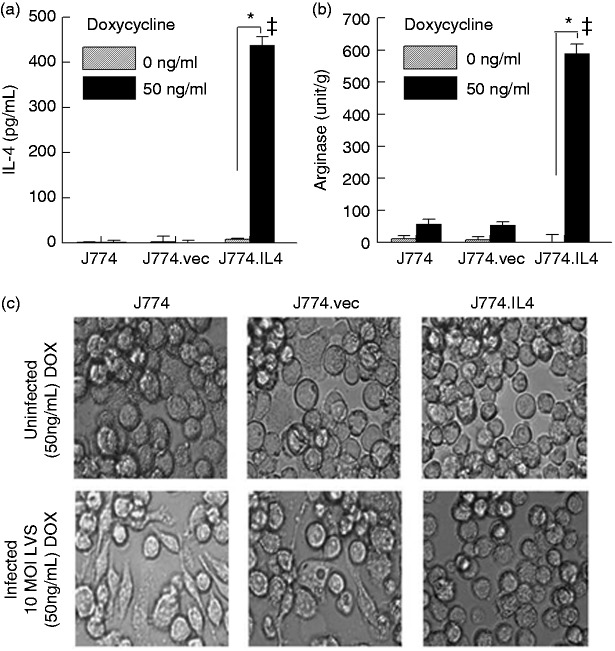
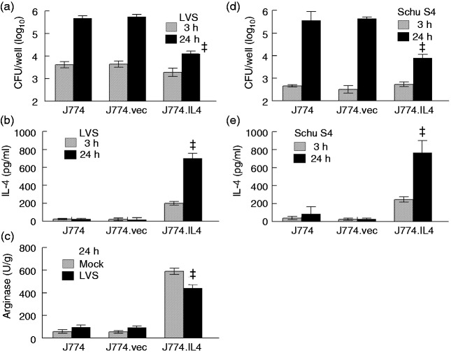
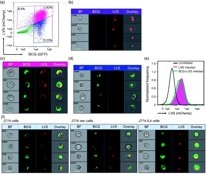
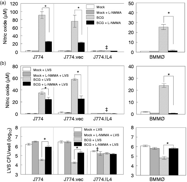
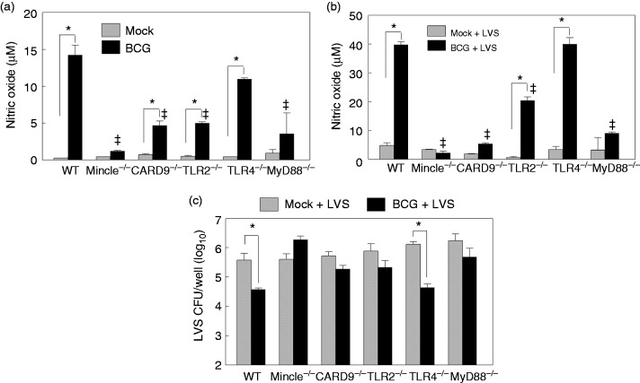
Macrophages are important innate immune cells that respond to microbial insults. In response to multi-bacterial infection, the macrophage activation state may change upon exposure to nascent mediators, which results in different bacterial killing mechanism(s). In this study, we utilized two respiratory bacterial pathogens, Mycobacterium bovis (Bacillus Calmette Guẻrin, BCG) and Francisella tularensis live vaccine strain (LVS) with different phagocyte evasion mechanisms, as model microbes to assess the influence of initial bacterial infection on the macrophage response to secondary infection. Non-activated (M0) macrophages or activated M2-polarized cells (J774 cells transfected with the mouse IL-4 gene) were first infected with BCG for 24-48 h, subsequently challenged with LVS, and the results of inhibition of LVS replication in the macrophages was assessed. BCG infection in M0 macrophages activated TLR2-MyD88 and Mincle-CARD9 signaling pathways, stimulating nitric oxide (NO) production and enhanced killing of LVS. BCG infection had little effect on LVS escape from phagosomes into the cytosol in M0 macrophages. In contrast, M2-polarized macrophages exhibited enhanced endosomal acidification, as well as inhibiting LVS replication. Pre-infection with BCG did not induce NO production and thus did not further reduce LVS replication. This study provides a model for studies of the complexity of macrophage activation in response to multi-bacterial infection.

Keywords: BCG; Francisella; IL-4; co-infection; macrophage.


Conflict of interest statement

The author(s) declared no potential conflicts of interest with respect to the research, authorship, and/or publication of this article.

Figures

Figure 1.
Figure 1.
Alternative activation of J774.IL-4 macrophages. J774, J774.vec or J774.IL4 cells were grown for 12 h in the presence (50 ng/ml) or absence of DOX. IL-4 secretion (a) in culture media was measured by ELISA, cellular arginase activity (b) was determined using the QuantiChrom Arginase Assay Kit, and cellular morphology (c) was visualized (400× phase contrast microscopy) 24 h after LVS inoculation (10 MOI). *P < 0.05 between indicated groups. ǂP < 0.05 between the indicated J774.IL4 and respective J774 and J774.vec groups. BCG: Bacillus Calmette Guẻrin; DOX: doxycycline; LVS: live vaccine strain.
Figure 2.
Figure 2.
F. tularensis replication in J774 cell lines. J774, J774.vec and J774.IL4 cells (5 × 105 per well) were incubated with doxycycline (50 ng/ml) for 12 h and subsequently infected with 5 × 106 LVS ((a) to (c)) or Schu S4 ((d) and (e)). Bacterial uptake and replication were measured 3 h and 24 h post-inoculation ((a) and (d)). Additionally, the IL-4 concentration in culture media was measured ((b) and (e)) and the cellular arginase activity was determined (C). ǂP < 0.05 between the indicated J774.IL4 and respective J774 and J774.vec groups. LVS: live vaccine strain.
Figure 3.
Figure 3.
Uptake of BCG and LVS by J774 macrophage-like cells. Untreated J774 cells ((a) to (e)) or 50 ng/ml doxycycline-treated J774, J774.vec and J774.IL4 cells (f) were seeded (5 × 105/well in 24-well plates) for 12 h followed by infection with GFP-expressing BCG (10 MOI) for 48 h, and subsequent inoculation with mCherry-expressing LVS (10 MOI). After 4 h incubation, co-infection with BCG and LVS was visualized and frequency analyzed using the Imagestream MKII (Amnis, EMD Millipore). (a) The dot-plot depicts the GFP and mCherry intensity of each cell and three gated cell populations: (b) mCherryhiGFPlow, (c) mCherryhiGFPhi and (d) mCherrylowGFPhi. The representative cell images of these three gated populations are shown in (b) LVS-infected J774 cells, (c) BCG–LVS co-infected cells (cellular localization of bacteria within this population was 93% using the Internalization Index analysis, IDEAS®) and (d) BCG-infected cells. (e) Uptake of mCherry LVS by J774 cells was comparable between LVS alone (pink area) and BCG + LVS co-infection (green line). (F) Shown are representative J774, J774.vec and J774.IL4 cells co-infected with BCG and LVS. BCG: Bacillus Calmette Guẻrin; BF: Bright Field; LVS: live vaccine strain.
Figure 4.
Figure 4.
BCG and LVS co-infection in J774 cell lines. J774 cell lines (5 × 105) were seeded in wells on cover slips and IL-4 expression induced with 50 ng/ml doxycycline for 12 h. Cells were then infected with either (a) LVS (5 × 106) or (b) BCG (5 × 106) for 24 h, followed by LVS (5 × 106). At 1 and 4 h post-LVS inoculation, cells were fixed, treated with either Dig or Sap, and stained for LVS. The cytosolic (LVS-Dig and BCG + LVS-Dig) and total intracellular (LVS-Sap and BCG + LVS-Sap) LVS quantifications were determined as described in the Materials and methods. Concurrently, J774 cell lines (5 × 105 per well) were induced with doxycycline, infected with BCG or mock treated with medium for 48 h, and infected with LVS. Uptake and replication of LVS were measured at 3 and 24 h post-LVS inoculation (c), and NO levels in culture medium were measured prior to and 24 h after LVS inoculation using the Griess reagent (d). *P < 0.05 between indicated groups. ǂP < 0.05 between the indicated J774.IL4 and respective J774 and J774.vec groups. BCG: Bacillus Calmette Guẻrin; Dig: digoxin; LVS: live vaccine strain; Sap: saponin.
Figure 5.
Figure 5.
NO-mediated control of LVS replication. J774, J774.vec and J774.IL-4 cells (1 × 106 per well) were induced with doxycycline (50 ng/ml, 12 h). BMMØs derived from C57BL/6 mice were infected with BCG (1 × 107) in the presence or absence of NO inhibitor L-NMMA, and NO levels in culture media measured 24 h post-inoculation (a). In a similar study, BMMØs and doxycycline-induced J774 cell lines were infected with BCG, or mock treated with medium in the presence or absence of L-NMMA for 48 h, and infected with LVS. NO concentration ((b), upper panel) and LVS replication ((b), lower panel) were measured 24 h post-LVS inoculation. *P < 0.05 between indicated groups. ǂP < 0.05 between the indicated J774.IL4 and respective J774 and J774.vec groups. BCG: Bacillus Calmette Guẻrin; BMMØ: bone marrow-derived macrophages; L-NMMA: NG-monomethyl-L-arginine acetate salt; LVS: live vaccine strain.
Figure 6.
Figure 6.
Innate signaling is required for BCG-mediated LVS inhibition during BCG–LVS superinfection. BMMØs prepared from WT C57BL6 and various gene-deficient mice, including Mincle, CARD9, TLR2, TLR4 and MyD88, were infected with BCG for 48 h and NO production was measured (a). Similarly prepared BMMØs were infected with LVS for 24 h with (BCG + LVS) or without (mock + LVS) prior BCG (48 h) infection, and the NO levels in culture medium (b) and viable LVS within macrophages (c) were analyzed. *P < 0.05 between indicated groups. ǂP < 0.05 between the indicated gene-deficiency and WT groups. BCG: Bacillus Calmette Guẻrin; BMMØ: bone marrow-derived macrophages; CARD9: caspase recruitment domain family member 9; LVS: live vaccine strain; WT, wild type.
Figure 7.
Figure 7.
Working model for BCG-mediated LVS inhibition during BCG–LVS superinfection. Shown in the proposed model are distinct LVS inhibition mechanisms following BCG–LVS superinfection in M0 J774/BMMØs and M2 J774.IL4 cells. In the M0 macrophage, BCG infection activates TLR2-MyD88 and Mincle-CARD9 signal pathways leading to NO production for subsequent LVS killing. In contrast, BCG has minimal effect on IL-4 mediated LVS killing in J774.IL4 M2 macrophages. BCG: Bacillus Calmette Guẻrin; BMMØ: bone marrow-derived macrophages; iNOS: inducible NO synthase; CARD9: caspase recruitment domain family member 9; LVS: live vaccine strain; M0: non-activated.

References

    1. Lijek RS, Weiser JN. Co-infection subverts mucosal immunity in the upper respiratory tract. Curr Opin Immunol 2012; 24: 417–423. - PMC - PubMed
    1. Bakaletz LO. Developing animal models for polymicrobial diseases. Nat Rev Microbiol 2004; 2: 552–568. - PMC - PubMed
    1. Arrevillaga G, Gaona J, Sanchez C, et al. Respiratory syncytial virus persistence in macrophages downregulates intercellular adhesion molecule-1 expression and reduces adhesion of non-typeable Haemophilus influenzae. Intervirology 2012; 55: 442–450. - PubMed
    1. Gomes MS, Paul S, Moreira AL, et al. Survival of Mycobacterium avium and Mycobacterium tuberculosis in acidified vacuoles of murine macrophages. Infect Immun 1999; 67: 3199–3206. - PMC - PubMed
    1. Draijer C, Robbe P, Boorsma CE, et al. Characterization of macrophage phenotypes in three murine models of house-dust-mite-induced asthma. Mediators Inflamm 2013; 2013: 632049–632049. - PMC - PubMed

Publication types

LinkOut - more resources