. 2018 Feb 26;19(1):25.
doi: 10.1186/s13059-018-1400-x.

i-GONAD: a robust method for in situ germline genome engineering using CRISPR nucleases

Affiliations

i-GONAD: a robust method for in situ germline genome engineering using CRISPR nucleases

Masato Ohtsuka et al. Genome Biol. .

Abstract

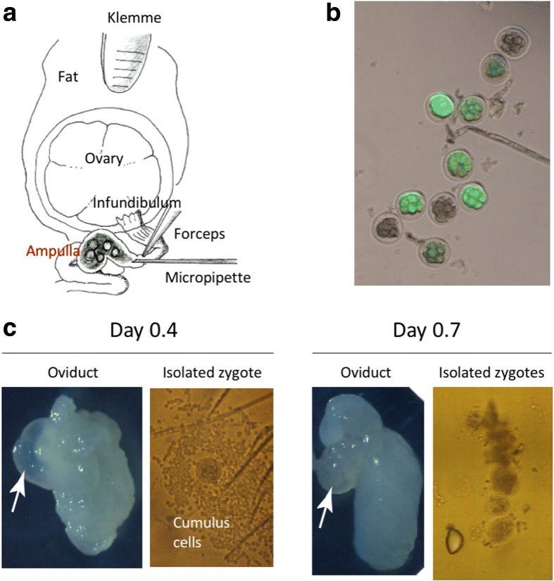
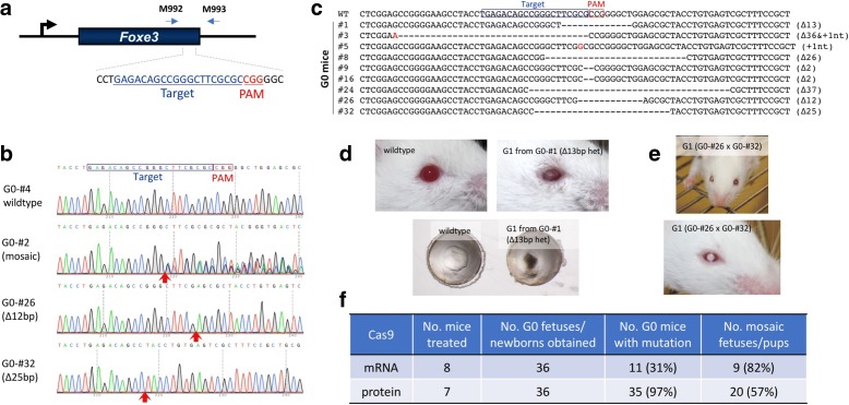
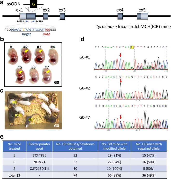
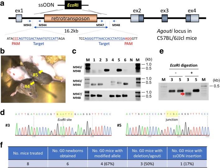
We present a robust method called improved-Genome editing via Oviductal Nucleic Acids Delivery (i-GONAD) that delivers CRISPR ribonucleoproteins to E0.7 embryos via in situ electroporation. The method generates mouse models containing single-base changes, kilobase-sized deletions, and knock-ins. The efficiency of i-GONAD is comparable to that of traditional microinjection methods, which rely on ex vivo handling of zygotes and require recipient animals for embryo transfer. In contrast, i-GONAD avoids these technically difficult steps, and it can be performed at any laboratory with simple equipment and technical expertise. Further, i-GONAD-treated females retain reproductive function, suggesting future use of the method for germline gene therapy.

Keywords: CRISPR; Easi-CRISPR; GONAD; In vivo electroporation; Knock-in; Long ssDNA; Transgenic mouse.


Conflict of interest statement

Ethics approval and consent to participate

All animal experiments performed were approved by the institutional protocols (Tokai University #154014, #165009, #171003; Hamamatsu University #2017062; and Shigei Medical Research Institute #17008).

Competing interests

MO, CBG, and MS have filed a provisional patent application relating to the work described in this manuscript. Tokai University and BEX Co. Ltd. applied for a patent describing the electroporation condition using CUY21Edit II on application number 2017–233100 (filed December 5, 2017). MO is an inventor of the patent.

Publisher’s Note

Springer Nature remains neutral with regard to jurisdictional claims in published maps and institutional affiliations.

Figures

Fig. 1
Fig. 1
Evaluation of earlier time points for performing GONAD. a Diagrammatic illustration showing the anatomical structures of ovary and oviduct and the surgical equipment used for GONAD procedure. A small amount of solution is injected by direct insertion of a glass micropipette through oviduct wall located at the region between the ampulla and infundibulum. Immediately after injection, in vivo electroporation is performed on the entire oviduct. b Detection of eGFP fluorescence in 8-cell to morula embryos after delivery of eGFP mRNA via GONAD procedure. The eGFP fluorescence in preimplantation embryos, isolated 2 days post GONAD procedure performed on naturally mated Jcl:MCH(ICR) female at 0.7 day of pregnancy. c Oviducts and zygotes dissected on days 0.4 (left panel) and 0.7 (right panel). Note that the oviduct dissected on day 0.4 exhibits swelling of the ampulla (arrow). The zygotes isolated from the day 0.4 ampulla are usually surrounded by thick layer of cumulus cells. These cells may hamper efficient uptake of exogenous nucleic acids/proteins injected intra-oviductally and subsequently electroporated. The oviduct dissected on day 0.7 exhibits shrinkage of the ampulla (arrow), and zygotes isolated from the day 0.7 ampulla have fewer cumulus cells, which will less likely hamper the uptake of exogenous nucleic acids/proteins upon electroporation
Fig. 2
Fig. 2
Creating gene-inactivated animal models using the GONAD method. a Schematic of the targeting strategy to inactivate Foxe3 gene and the primer set used for genotyping. b Direct sequencing results of polymerase chain reaction (PCR) products amplified from the founder (G0) mice with the primer set shown in a. The red arrows below the electropherogram show the region with indel mutations. c Mutated Foxe3 alleles in the G0 mice. The changes in the nucleotide sequence are shown in red, and the type of changes (insertions +Xnt, or deletions Δ) is indicated on the right side of the sequences. d and e Cataract phenotypes in the G1 mice. f Efficiencies of Foxe3 gene editing. CRISPR components used were either Cas9 mRNA/sgRNA or Cas9 protein/CRISPR RNA (crRNA)/trans-activating crRNA (tracrRNA) (see Additional file 1: Table S1 for details)
Fig. 3
Fig. 3
Creating small genetic change animal models using the i-GONAD method. Restoration of Tyr gene of albino Jcl:MCH(ICR) mice by single-stranded oligo donor (ssODN)-based knock-in with the i-GONAD method. a Schematic to show rescue of Tyr gene mutation. The target region containing the guide sequence and the genotyping primer binding sites are shown. b Representative E14.5 litter showing Tyr rescued G0 fetuses. The pigmented eyes of the fetuses are indicated by yellow arrows. c Representative Tyr rescued G0 mouse litters obtained from #5 female mouse in Additional file 1: Table S2. G0 mice indicated in # numbers (shown in yellow) were used for germline transmission analysis (see details in Additional file 1: Figure S2). d Direct sequencing results of PCR products amplified from the G0 fetuses in b. The positions for mutated/corrected nucleotides are indicated by red arrows. e Efficiencies of Tyr gene editing. Electroporators from three different suppliers were used (see Additional file 1: Table S2 for details)
Fig. 4
Fig. 4
Creating large deletion using the i-GONAD method. a Schematic diagram showing deletion of 16.2-kb sequence consisting of retrotransposon in the C57BL/6JJcl mouse genome, to restore agouti phenotype. The target sequences and genotyping primers are shown. ssODN containing EcoRI site at the middle of the sequence was used. b Representative mice showing rescued agouti phenotype (indicated by yellow arrows). These mice were recovered through caesarean section and nursed by Jcl:MCH(ICR) foster mother with her own pups. c Genotyping analyses. Expected fragment sizes: M943/M948 = 290 or 295 bp (ssODN knock-in), M943/M944 = 337 bp, M947/M948 = 477 bp. d Direct sequencing results of PCR products amplified from the G0 mice. The position of junctional sequences is indicated by yellow rectangles. e EcoRI digestion of PCR products amplified from G0 mice (G0-#3 and -#5) with the M943/M948 primer set. Red arrow indicates digested fragment. f Efficiencies of agouti gene editing (see Additional file 1: Table S4 for details)
Fig. 5
Fig. 5
Generation of reporter knock-in mice using the i-GONAD method. a Schematic diagram showing insertion of T2A-mCitrine cassette into Pitx3 locus. The target sequence and the genotyping primer sets are shown. A 925-base-long ssDNA synthesized by ivTRT method was used as the donor DNA. b mCitrine fluorescence in fetus collected at E12.5. The eye of the fetus is enlarged as an inset. c Example of genotyping analysis of knock-in G0 fetuses. Expected fragment sizes: M1035/M390 = 948 bp, M389/M1036 = 956 bp, M389/PP226 = 809 bp. N negative control, M size marker. d Representative sequencing chromatogram showing 5′ and 3′ junctional regions of the inserted cassette. The junctional sequences showing insertion derived from G0-#1 in c are shown. Red arrows indicate junctions between the arms and the genomic sequences. e Genome editing efficiency of the Pitx3 locus by the i-GONAD method

Comment in

References

    1. Harms DW, Quadros RM, Seruggia D, Ohtsuka M, Takahashi G, Montoliu L, et al. Mouse genome editing using the CRISPR/Cas system. Curr Protoc Hum Genet. 2014;83:15.7.1–15.7.27. - PMC - PubMed
    1. Wang H, Yang H, Shivalila CS, Dawlaty MM, Cheng AW, Zhang F, et al. One-step generation of mice carrying mutations in multiple genes by CRISPR/Cas-mediated genome engineering. Cell. 2013;153:910–918. doi: 10.1016/j.cell.2013.04.025. - DOI - PMC - PubMed
    1. Gurumurthy CB, Grati M, Ohtsuka M, Schilit SLP, Quadros RM, Liu XZ. CRISPR: a versatile tool for both forward and reverse genetics research. Hum Genet. 2016;135:971–976. doi: 10.1007/s00439-016-1704-4. - DOI - PMC - PubMed
    1. Hashimoto M, Yamashita Y, Takemoto T. Electroporation of Cas9 protein/sgRNA into early pronuclear zygotes generates non-mosaic mutants in the mouse. Dev Biol. 2016;418:1–9. doi: 10.1016/j.ydbio.2016.07.017. - DOI - PubMed
    1. Kaneko T, Mashimo T. Simple genome editing of rodent intact embryos by electroporation. PLoS One. 2015;10:e0142755. doi: 10.1371/journal.pone.0142755. - DOI - PMC - PubMed

Publication types