

Case Reports
doi: 10.1128/AAC.00172-18.
Print 2018 May.
In Situ Validation of the Endothelial Cell Receptor GRP78 in a Case of Rhinocerebral Mucormycosis
Affiliations
- PMID: 29483124
- PMCID: PMC5923111
- DOI: 10.1128/AAC.00172-18
Item in Clipboard
Case Reports
In Situ Validation of the Endothelial Cell Receptor GRP78 in a Case of Rhinocerebral Mucormycosis
Antimicrob Agents Chemother.
.
No abstract available
Keywords: GRP78; endothelium invasion; glucose-regulated protein; mucormycosis.
Figures
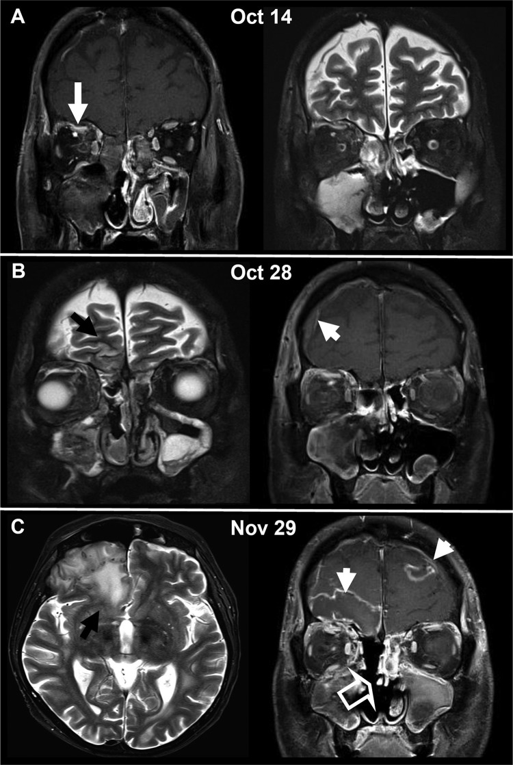
Cranial MRI illustrating the progression of cerebral and orbital involvement of mucormycosis with R. oryzae over a time span of 6 weeks. (A) Initial scan showing the obliterated maxillary and ethmoidal sinuses on the right side and the orbital phlegmonous infection as well as a small subperiosteal abscess under the orbital roof (white arrow). (B) Progressive disease 2 weeks after the initial scan with diffuse edema and reticular contrast enhancement in the right retroorbital space. Signs of intracranial per continuitatem dissemination with frontobasal cortical edema (black arrow) and meningeal contrast enhancement (white arrowhead) are evident. Despite surgery, acute inflammatory mucosal swelling of the right maxilla persisted. (C) With further progression 6 weeks after the initial scan, the patient exhibited massive perifocal edema in the frontobasal brain parenchyma (black arrow), a mild frontal midline shift, and a typical MRI appearance of an intracranial abscess with central necrosis and intense peripheral contrast enhancement (white arrowheads). Progressive inflammatory changes in the left frontal and the left maxillary sinus after resection of the right ethmoid cellulae and the right lower and middle concha (open arrow) were observed.
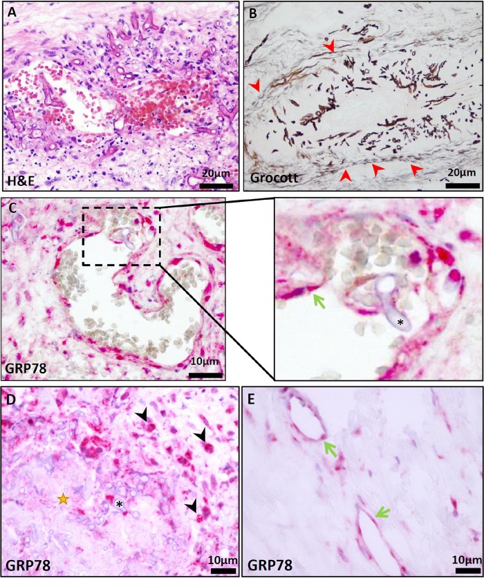
(A) H&E staining of the resected sinoid mucosa presented angioinvasive mucormycosis, causing thrombosis and widespread necrotic areas (scale bar, 20 μm). (B) Grocott staining confirmed angioinvasive infiltration. Red arrowheads highlight a vessel wall infiltrated with R. oryzae organisms (scale bar, 20 μm). (C) Immunohistochemical GRP78 staining (GRP78/BiP, C50B12, 1:200; Cell Signaling Technology) demonstrated an intense expression on infiltrated endothelial cells (scale bar, 10 μm). (Inset in panel C) Higher magnification. The green arrow marks endothelial cells close to R. oryzae (*). (D) Black arrowheads highlight interstitial GRP78-positive macrophages demarking necrotic areas (star) infiltrated with R. oryzae (*) (scale bar, 10 μm). (E) GRP78 staining of endothelial cells (green arrows) not affected by R. oryzae in the same section demonstrates much lower expression levels (scale bar, 10 μm).
References
Publication types
MeSH terms
Substances
Grants and funding
LinkOut - more resources
Full Text Sources
Other Literature Sources
Miscellaneous
