. 2018 Feb 1;32(3-4):271-282.
doi: 10.1101/gad.310672.117. Epub 2018 Feb 26.

C. elegans ADARs antagonize silencing of cellular dsRNAs by the antiviral RNAi pathway

Affiliations

C. elegans ADARs antagonize silencing of cellular dsRNAs by the antiviral RNAi pathway

Daniel P Reich et al. Genes Dev. .

Abstract

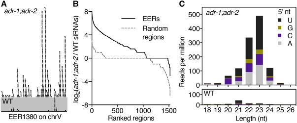
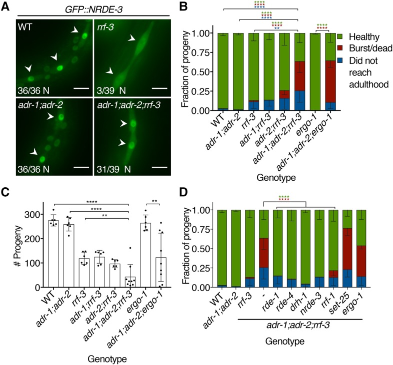
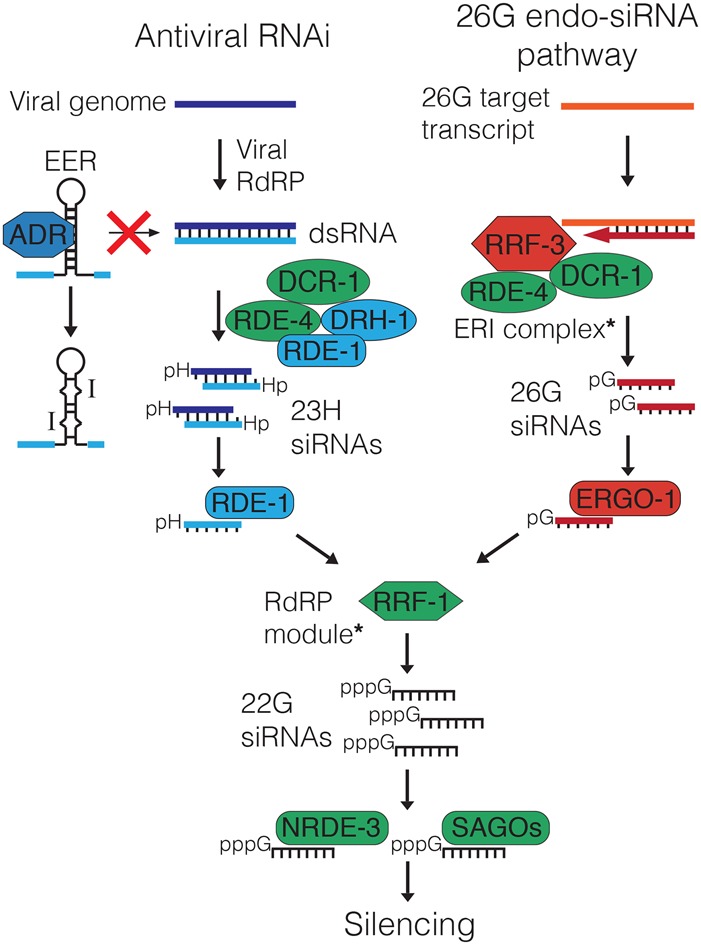
Cellular dsRNAs are edited by adenosine deaminases that act on RNA (ADARs). While editing can alter mRNA-coding potential, most editing occurs in noncoding sequences, the function of which is poorly understood. Using dsRNA immunoprecipitation (dsRIP) and RNA sequencing (RNA-seq), we identified 1523 regions of clustered A-to-I editing, termed editing-enriched regions (EERs), in four stages of Caenorhabditis elegans development, often with highest expression in embryos. Analyses of small RNA-seq data revealed 22- to 23-nucleotide (nt) siRNAs, reminiscent of viral siRNAs, that mapped to EERs and were abundant in adr-1;adr-2 mutant animals. Consistent with roles for these siRNAs in silencing, EER-associated genes (EAGs) were down-regulated in adr-1;adr-2 embryos, and this was dependent on associated EERs and the RNAi factor RDE-4. We observed that ADARs genetically interact with the 26G endogenous siRNA (endo-siRNA) pathway, which likely competes for RNAi components; deletion of factors required for this pathway (rrf-3 or ergo-1) in adr-1;adr-2 mutant strains caused a synthetic phenotype that was rescued by deleting antiviral RNAi factors. Poly(A)+ RNA-seq revealed EAG down-regulation and antiviral gene induction in adr-1;adr-2;rrf-3 embryos, and these expression changes were dependent on rde-1 and rde-4 Our data suggest that ADARs restrict antiviral silencing of cellular dsRNAs.

Keywords: Dicer; RNA editing; deaminase; self–nonself; siRNA.


Figures

Figure 1.
Figure 1.
EER abundance, but not editing, changes during development. (E) Early; (L) late; (Y) young. (A) The number of EERs defined from each group of data sets. (B) Venn diagram of EERs defined in each developmental stage. (C) Heat map of relative abundance in input RNA-seq samples for the 250 EERs with the greatest number of edited windows. (D) Distribution of mean EER abundance in each stage and treatment. (****) P < 0.0001, Mann-Whitney U-test. (E) Fraction of all EER-editing sites that appeared as guanosines in each stage and treatment. While individual sites ranged from 1% to 99% edited, all sites together were ∼15% edited in each sample. Error bars show standard deviation (SD) of three biological replicates. (*) P < 0.05, Student's t-test.
Figure 2.
Figure 2.
EER-23H siRNAs are abundant in adr-1;adr-2 double mutants. (A) Genome browser view of 5′P-dependent small RNA-seq reads from mixed-stage wild-type (WT) and adr-1(tm668);adr-2(ok735) mutant animals mapping sense to EER1380. (B) EER-23H siRNA enrichment in adr-1(tm668);adr-2(ok735) mutants. Plots show the log2 ratio of siRNA abundance in adr-1;adr-2 mutants over wild type for EERs (black solid line) and control regions (gray dashed line). (C) Analysis of 5′ nucleotide and length distribution of all EER-23H siRNAs from adr-1;adr-2 mutant and wild-type animals.
Figure 3.
Figure 3.
EAG expression decreases in adr-1(uu49);adr-2(uu28) embryos in an RNAi- and EER-dependent manner. Expression of EAGs in embryos (A; n ≥ 8) and L3 larvae (B; n = 5) of four genotypes, measured by qRT–PCR. Expression levels for three EAGs in embryos (C; n ≥ 6) and L3 larvae (D; n = 5) in strains where each EAG's sole EER was deleted by CRISPR (ΔEER). All panels show expression as mean. Error bars show SD. (*) P < 0.05; (**) P < 0.01; (***) P < 0.001; (****) P < 0.0001, Student's t-test.
Figure 4.
Figure 4.
The 26G endo-siRNA pathway genetically interacts with ADARs. (A) GFP::NRDE-3 visualized in L3 larvae seam cells (arrowheads) of the indicated mutant genotypes. Numbers in the bottom left of each panel report the fraction of worms with nuclear-enriched (N) GFP::NRDE-3 in seam cells. Bar, 10 µm. (B) Bursting assay shows the fate of embryos laid by each genotype 5 d after egg lay. Error bars show SD. n ≥ 6 assays. (**) P < 0.01; (****) P < 0.0001. Asterisk colors show categories compared by two-way ANOVA with Tukey's multiple comparisons correction. (C) Average brood size for each genotype in B, with individual broods shown as dots. Error bars show SD. n ≥ 6 assays. (**) P < 0.01; (****) P < 0.0001, Student's t-test. (D) Developmental fates, as in B, of adr-1(uu49);adr-2(uu28);rrf-3(uu56) mutant strains with additional mutations in genes encoding RNAi-related factors. Error bars show SD. n ≥ 6 assays. (****) P < 0.0001. Asterisk colors show categories compared by two-way ANOVA with Tukey's multiple comparisons correction.
Figure 5.
Figure 5.
EAGs and Orsay virus-induced genes are misregulated when ADARs and the 26G pathway are disrupted. (A) Tukey box plots show distributions of log2(expression fold change compared with wild type) for EAGs in each mutant genotype analyzed by RNA-seq. (*) P < 0.05; (****) P < 0.0001, Mann-Whitney U-test. (B) Venn diagram showing the overlap between differentially expressed genes (DEGs) up-regulated and down-regulated in adr-1(uu49);adr-2(uu28);rrf-3(uu56) mutants compared with wild type as well as EAGs expressed in RNA-seq samples (>10 reads total). Tukey box plots as in A show expression fold change in mutant genotypes for significantly down-regulated EAGs (down) (C), significantly up-regulated EAGs (up) (D), and EAGs not significantly changed (NS) (E) in adr-1;adr-2;rrf-3 mutant embryos relative to wild type. Adjusted P-value cutoff was 0.05. (ns) P > 0.05; (*) P < 0.05; (**) P < 0.01; (****) P < 0.0001, Mann-Whitney U-test. (F) Genes analyzed by poly(A)+ RNA-seq are plotted by log2(expression fold change compared with wild type) against −10log10(adjusted P-value) in adr-1;adr-2;rrf-3 mutants compared with wild type (i.e., higher y-values indicate more significant differences). The horizontal dotted line designates the adjusted P-value cutoff of 0.05 used to define DEGs. (G) Tukey box plots as in A showing Orsay-induced gene expression fold change in mutants. (**) P < 0.01; (****) P < 0.0001, Mann-Whitney U-test.
Figure 6.
Figure 6.
ADARs and the 26G pathway prevent antiviral RNAi-mediated silencing of self dsRNAs. During viral infection, viral replication generates dsRNAs that are processed into 23H siRNAs by a complex of DCR-1, RDE-1, RDE-4, and DRH-1. RDE-1 binds 23H siRNAs and stimulates 22G siRNA production by RRF-1. 22G siRNAs bind NRDE-3 and SAGO Argonautes to effect silencing. ADARs bind and edit EERs to prevent recognition as viral dsRNA and processing to 23H siRNAs by the antiviral DCR-1 complex. In the 26G pathway, the ERI complex (containing RRF-3, DCR-1, and RDE-4) generates 26G siRNAs, which bind ERGO-1, promote 22G siRNA synthesis, and silence targets through NRDE-3 and SAGO proteins. Thus, absent viral infection, antiviral RNAi is kept inactive by ADARs binding and editing self dsRNAs and by 26G pathway sequestration of common RNAi factors (green). For simplicity, we show only factors relevant to this study (see Supplemental Fig. S1); complexes containing additional components are noted with asterisks.

Comment in

  • A rADAR defense against RNAi.
    Pasquinelli AE. Pasquinelli AE. Genes Dev. 2018 Feb 1;32(3-4):199-201. doi: 10.1101/gad.313049.118. Genes Dev. 2018. PMID: 29491134 Free PMC article.

References

    1. Aoki K, Moriguchi H, Yoshioka T, Okawa K, Tabara H. 2007. In vitro analyses of the production and activity of secondary small interfering RNAs in C. elegans. EMBO J 26: 5007–5019. - PMC - PubMed
    1. Ashe A, Belicard T, Le Pen J, Sarkies P, Frezal L, Lehrbach NJ, Felix MA, Miska EA. 2013. A deletion polymorphism in the Caenorhabditis elegans RIG-I homolog disables viral RNA dicing and antiviral immunity. Elife 2: e00994. - PMC - PubMed
    1. Batista PJ, Ruby JG, Claycomb JM, Chiang R, Fahlgren N, Kasschau KD, Chaves DA, Gu W, Vasale JJ, Duan S, et al. 2008. PRG-1 and 21U-RNAs interact to form the piRNA complex required for fertility in C. elegans. Mol Cell 31: 67–78. - PMC - PubMed
    1. Billi AC, Fischer SE, Kim JK. 2014. Endogenous RNAi pathways in C. elegans. WormBook (ed. The C. elegans Research Community). WormBook 10.1895/wormbook.1.170.1, http://www.wormbook.org. - DOI - PMC - PubMed
    1. Blango MG, Bass BL. 2016. Identification of the long, edited dsRNAome of LPS-stimulated immune cells. Genome Res 26: 852–862. - PMC - PubMed

Publication types

MeSH terms