. 2018 Feb 26;8(1):3609.
doi: 10.1038/s41598-018-21517-4.

MicroRNA Contents in Matrix Vesicles Produced by Growth Plate Chondrocytes are Cell Maturation Dependent

Affiliations

MicroRNA Contents in Matrix Vesicles Produced by Growth Plate Chondrocytes are Cell Maturation Dependent

Zhao Lin et al. Sci Rep. .

Abstract

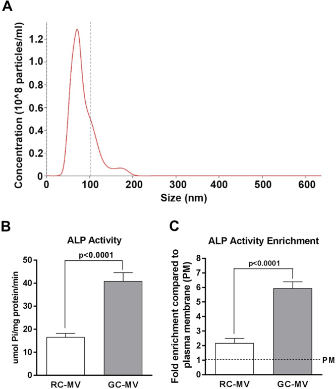
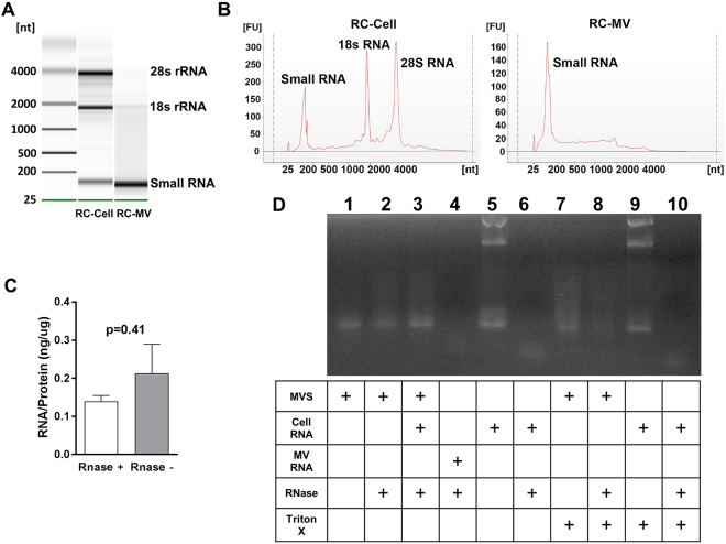
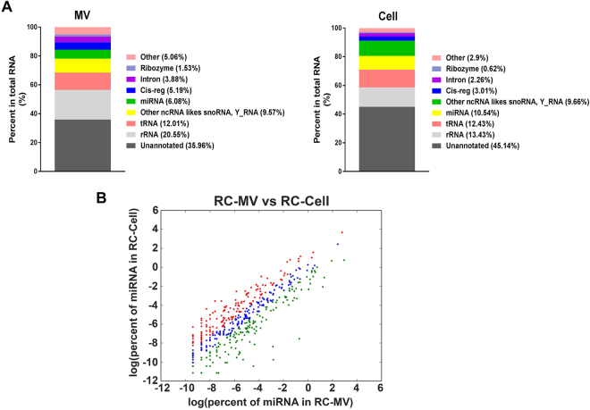
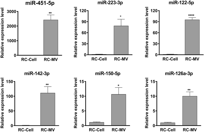
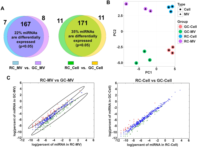
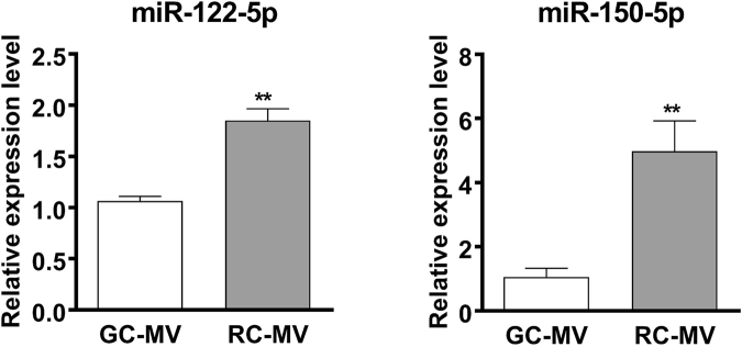
Chondrocytes at different maturation states in the growth plate produce matrix vesicles (MVs), membrane organelles found in the extracellular matrix, with a wide range of contents, such as matrix processing enzymes and receptors for hormones. We have shown that MVs harvested from growth zone (GC) chondrocyte cultures contain abundant small RNAs, including miRNAs. Here, we determined whether RNA also exists in MVs produced by less mature resting zone (RC) chondrocytes and, if so, whether it differs from the RNA in MVs produced by GC cells. Our results showed that RNA, small RNA specifically, was present in RC-MVs, and it was well-protected from RNase by the phospholipid membrane. A group of miRNAs was enriched in RC-MVs compared RC-cells, suggesting that miRNAs are selectively packaged into MVs. High throughput array and RNA sequencing showed that ~39% miRNAs were differentially expressed between RC-MVs and GC-MVs. Individual RT-qPCR also confirmed that miR-122-5p and miR-150-5p were expressed at significantly higher levels in RC-MVs compared to GC-MVs. This study showed that growth plate chondrocytes at different differentiation stages produce different MVs with different miRNA contents, further supporting extracellular vesicle miRNAs play a role as "matrisomes" that mediate the cell-cell communication in cartilage and bone development.


Conflict of interest statement

The authors declare no competing interests.

Figures

Figure 1
Figure 1
Characterization of RC-MVs. (A) Nanosight was used to measure the size of RC-MVs, which showed an average diameter around 80 nm. (B) Alkaline phosphatase (ALP) specific activity was compared between RC-MVs and GC-MVs. GC cells produced MVs with higher ALP specific activity than RC cells (n = 5 or 6). (C) In RC cell cultures, less ALP activity enrichment was seen in MVs compared to that in GC cell cultures (n = 5 or 6). PM=plasma membrane.
Figure 2
Figure 2
RNA exists in RC-MVs. (A) MV RNA was compared to cell RNA. In a 2% agarose gel, RC-MV RNA was heterogeneous in size but contained little or no large ribosomal RNA species (18S- and 28S-rRNA) compared to parent cells. Enriched small RNAs were observed in MVs. (B) A similar pattern was observed in the Bioanalyzer. (C) RC-MVs were treated by RNase A before RNA extraction by TRIzol. No significant differences in the RNA:protein ratios of MVs with or without RNase pretreatment (n = 6) were observed. (D) RNase was not able to digest the RNA component in MVs. However, it was able to digest the total cell RNA. When the MVs were pre-treated with the membrane detergent Triton X-100, RNA degradation was observed, suggesting that RNA in RC-MVs is protected by the intact lipid membrane.
Figure 3
Figure 3
The profiles of small RNAs in RC-MVs and parent cells. (A) Complex populations of coding and non-coding small RNAs were found in RC-MVs in proportions that were distinct from those in the RC cells. (B) miRNAs in RC-MVs and RC-cells identified by RNA-seq were plotted. Red dots represent miRNAs with at least a 2-fold expression level increase in MVs than those in cells. Green dots represent miRNAs with more than a 2-fold expression level increase in cells than that in MVs. Blue indicates the overlap between these two groups. A large number of miRNAs were differentially expressed between RC-MVs and RC-cells.
Figure 4
Figure 4
Validation of the miRNA enriched in RC-MVs compared with parent cells. Six miRNAs (miR-451-5p, miR-223-3p, miR-122-5p, miR-142-3p, miR-150-5p, miR-126a-3p) were selected for RT-qPCR assays. Consistent with previous array results, the expression of all these miRNAs were significantly higher in MVs compared to cells. These results suggest that certain miRNAs are selectively exported into MVs. *p < 0.05 in t-test. **p < 0.01 in t-test. ****p < 0.0001 in t-test (n = 3).
Figure 5
Figure 5
Distinct differences in the miRNAs between RC-MVs and GC-MVs. (A) Different miRNAs patterns were identified from PCR-array between two maturation status of chondrocytes, both in MVs and the parent cells. (B) Principal component analysis (PCA) of the miRNA PCR-array data for all the four different types of samples: RC-MVs, RC-cells, GC-MVs and GC-cells. The differences among these samples were clear, as larger difference showed on the x-axis direction compared to that on the y-axis. This suggests that the differences between MVs and cells were larger than between two different cell maturation stages. (C) Left panel: miRNAs in RC-MVs and GC-MVs identified by RNA-seq were plotted. Red dots represent miRNAs with at least a 2-fold expression level increase in GC-MVs than in RC-MVs. Green dots represent miRNAs with a more than 2-fold expression level increase in RC-MVs than in GC-MVs. Blue indicates the overlap between these two groups. A large number of miRNAs were differentially expressed between RC-MVs and GC-MVs. Right panel: similar comparison was done between RC-cells and GC-cells. Interestingly, the difference between RC-cells and GC-cells was not as large as that was seen between RC-MVs and GC-MVs.
Figure 6
Figure 6
RT-qPCR was used to further validate the differences in miRNA expression between RC-MVs and GC-MVs. miR-122-5p and miR-150-5p had significantly higher expression levels in RC-MVs than GC-MVs, suggesting the miRNAs contents in MVs are associated with the cell maturation status. **p < 0.01 in t-test. (n = 6).

Similar articles

Cited by

References

    1. Kronenberg HM. Developmental regulation of the growth plate. Nature. 2003;423:332–336. doi: 10.1038/nature01657. - DOI - PubMed
    1. Anderson HC. Vesicles associated with calcification in the matrix of epiphyseal cartilage. J. Cell Biol. 1969;41:59–72. doi: 10.1083/jcb.41.1.59. - DOI - PMC - PubMed
    1. Boyan BD, Schwartz Z, Swain LD, Carnes DL, Jr, Zislis T. Differential expression of phenotype by resting zone and growth region costochondral chondrocytes in vitro. Bone. 1988;9:185–194. doi: 10.1016/8756-3282(88)90008-7. - DOI - PubMed
    1. Dean DD, et al. Matrix vesicles are enriched in metalloproteinases that degrade proteoglycans. Calcif. Tissue Int. 1992;50:342–349. doi: 10.1007/BF00301632. - DOI - PubMed
    1. Dean DD, Boyan BD, Muniz OE, Howell DS, Schwartz Z. Vitamin D metabolites regulate matrix vesicle metalloproteinase content in a cell maturation-dependent manner. Calcif. Tissue Int. 1996;59:109–116. doi: 10.1007/s002239900096. - DOI - PubMed

Publication types