. 2018 Feb 26;8(1):3637.
doi: 10.1038/s41598-018-21804-0.

Characterization of the quinol-dependent nitric oxide reductase from the pathogen Neisseria meningitidis, an electrogenic enzyme

Affiliations

Characterization of the quinol-dependent nitric oxide reductase from the pathogen Neisseria meningitidis, an electrogenic enzyme

Nathalie Gonska et al. Sci Rep. .

Abstract

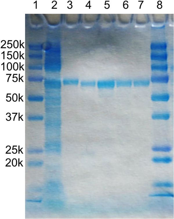
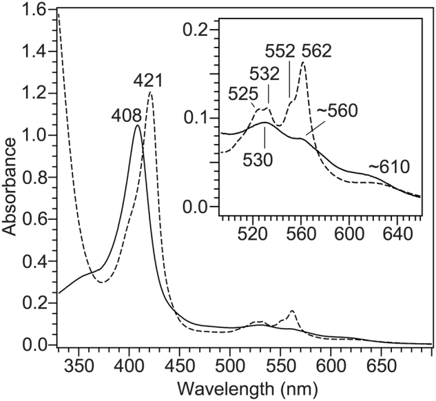
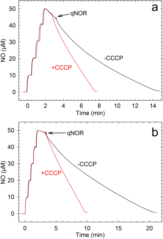
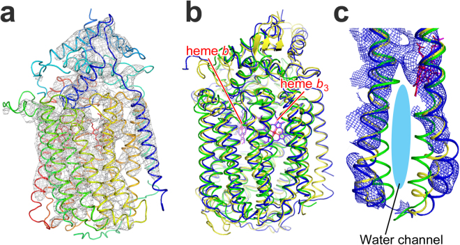
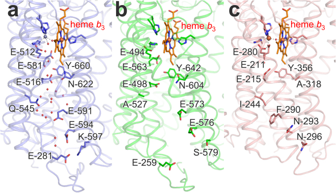
Bacterial nitric oxide reductases (NORs) catalyse the reduction of NO to N2O and H2O. NORs are found either in denitrification chains, or in pathogens where their primary role is detoxification of NO produced by the immune defense of the host. Although NORs belong to the heme-copper oxidase superfamily, comprising proton-pumping O2-reducing enzymes, the best studied NORs, cNORs (cytochrome c-dependent), are non-electrogenic. Here, we focus on another type of NOR, qNOR (quinol-dependent). Recombinant qNOR from Neisseria meningitidis, a human pathogen, purified from Escherichia coli, showed high catalytic activity and spectroscopic properties largely similar to cNORs. However, in contrast to cNOR, liposome-reconstituted qNOR showed respiratory control ratios above two, indicating that NO reduction by qNOR was electrogenic. Further, we determined a 4.5 Å crystal structure of the N. meningitidis qNOR, allowing exploration of a potential proton transfer pathway from the cytoplasm by mutagenesis. Most mutations had little effect on the activity, however the E-498 variants were largely inactive, while the corresponding substitution in cNOR was previously shown not to induce significant effects. We thus suggest that, contrary to cNOR, the N. meningitidis qNOR uses cytoplasmic protons for NO reduction. Our results allow possible routes for protons to be discussed.


Conflict of interest statement

The authors declare no competing interests.

Figures

Figure 1
Figure 1
SDS-PAGE of N. meningitidis qNOR samples during purification. Conditions: c-PAGEL (Atto corporation), 12.5% gel was used with Tris-glycine running buffer. Lanes 1 and 8: marker with molecular sizes as indicated, 2: solubilized membranes, 3: Ni-NTA purified qNOR, 4: HIS-tag cleaved qNOR, 5–7: SEC fractions.
Figure 2
Figure 2
Visible absorption spectra of wild-type N. meningitidis qNOR. The spectra shown are oxidized state (solid curve) and dithionite-reduced state (broken curve). The sample is in 50 mM Tris-HCl pH 8.0, 150 mM NaCl, and 0.1% DM. The reduced qNOR sample was prepared by the addition of an excess amount of dithionite to the oxidized enzyme under N2 atmosphere.
Figure 3
Figure 3
High frequency region of resonance Raman spectra of wild-type qNOR from N. meningitidis. Traces shown are (a) oxidized and (b) dithionite-reduced qNOR. The spectra were obtained with excitation at 413.1 nm. The qNOR concentration was adjusted to 20–40 µM in 50 mM Tris-HCl pH 8.0, 150 mM NaCl, and 0.1% DM. The reduced form was prepared by addition of an excess amount of dithionite under N2 atmosphere.
Figure 4
Figure 4
NO-reduction activity of N. meningitidis qNOR with MD/DTT (1 mM/5 mM, red trace) and PMS/Asc (10 µM/6 mM, black trace). Conditions: 20 mM K+-HEPES (pH 7.4), 100 mM KCl, 0.05% DDM, 10 mM glucose, 100 U/mL catalase, 10 U/mL glucose oxidase. About 5 times higher concentration of qNOR was used with PMS/Asc (~70 nM; black trace, compared to ~14 nM; red trace). NO was added in 5 consecutive steps (in total 50 µM); all additions except qNOR (addition indicated) were made before adding NO.
Figure 5
Figure 5
The effect of uncouplers on the activity of qNOR in liposomes. a: With MD/DTT and b: with Asc/PMS. Conditions: 20 mM K+-HEPES (pH 7.4), 100 mM KCl, 10 mM glucose, 100 U/mL catalase, 10 U/mL glucose oxidase, (+10 µM CCCP for red traces). All additions except qNOR were made before NO addition. qNOR in liposomes (40 nM) was added where indicated.
Figure 6
Figure 6
Crystal structure of N. meningitidis qNOR at a resolution 4.5 Å. (a) Overall structure with 2FoFc electron density map contoured at 2.0σ (gray mesh). (b) Superposition of the Cα traces of G. stearothermophilus qNOR (PDB ID: 3AYF, yellow ribbon) and P. aeruginosa cNOR (PDB ID: 3O0R, green ribbon) onto that of NmqNOR (blue ribbon). The root-mean-square deviations of the Cα atoms are 1.19 and 2.23 Å for NmqNOR-GsqNOR and NmqNOR-cNOR, respectively. Heme b and b3 in N. meningitidisG. stearothermophilus qNORs and cNOR are shown by red, magenta and blue sticks, respectively. (c) The water channel region in the superposed structure. The ribbon colors correspond to panel (b). Blue mesh represents a composite omit map of NmqNOR contoured at 1.5σ.
Figure 7
Figure 7
Structure of the water channel region in G. stearothermophilus qNOR (pdb ID 3AYF) (a), the corresponding region in the low-resolution structure of N. meningitidis qNOR (b) compared also to P. aeruginosa cNOR (pdb ID 3O0R) (c). Although the orientation of the side-chains of the amino acid residues could not be determined in the low resolution structure of NmqNOR, the residues that correspond to those in GsqNOR are modeled for positional comparison. Water molecules observed in the X-ray crystal structure of GsqNOR are shown as red spheres. The amino acid alignment (Supplementary Fig. S1) shows that E-281 in GsqNOR is replaced by a Thr (−255) in NmqNOR, but there is a Glu (−259) in a corresponding spatial location. Note that the iron in the FeB site (dark red sphere) in the cNOR structure (c) is replaced by a Zn ion (grey sphere) in GsqNOR, but that this site is presumably occupied by Fe in NmqNOR as described in Results. Figure made using the Pymol program.

References

    1. Tosha T, Shiro Y. Crystal structures of nitric oxide reductases provide key insights into functional conversion of respiratory enzymes. IUBMB Life. 2013;65:217–226. doi: 10.1002/iub.1135. - DOI - PubMed
    1. Pereira MM, Santana M, Teixeira M. A novel scenario for the evolution of haem-copper oxygen reductases. Biochim. Biophys. Acta. 2001;1505:185–208. doi: 10.1016/S0005-2728(01)00169-4. - DOI - PubMed
    1. Lee HJ, Reimann J, Huang Y, Ädelroth P. Functional proton transfer pathways in the heme-copper oxidase superfamily. Biochim. Biophys. Acta. 2012;1817:537–544. doi: 10.1016/j.bbabio.2011.10.007. - DOI - PubMed
    1. Sharma V, Wikström M. The role of the K-channel and the active-site tyrosine in the catalytic mechanism of cytochrome c oxidase. Biochim. Biophys. Acta. 2016;1857:1111–1115. doi: 10.1016/j.bbabio.2016.02.008. - DOI - PubMed
    1. Bell LC, Richardson DJ, Ferguson SJ. Identification of nitric oxide reductase activity in Rhodobacter capsulatus: the electron transport pathway can either use or bypass both cytochrome c2 and the cytochrome bc1 complex. J. Gen. Microbiol. 1992;138:437–443. doi: 10.1099/00221287-138-3-437. - DOI - PubMed

Publication types

MeSH terms

LinkOut - more resources