. 2018 Feb 26;9(1):815.
doi: 10.1038/s41467-018-03156-5.

Sculpting nanoparticle dynamics for single-bacteria-level screening and direct binding-efficiency measurement

Affiliations

Sculpting nanoparticle dynamics for single-bacteria-level screening and direct binding-efficiency measurement

Y Z Shi et al. Nat Commun. .

Erratum in

Abstract

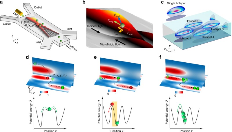
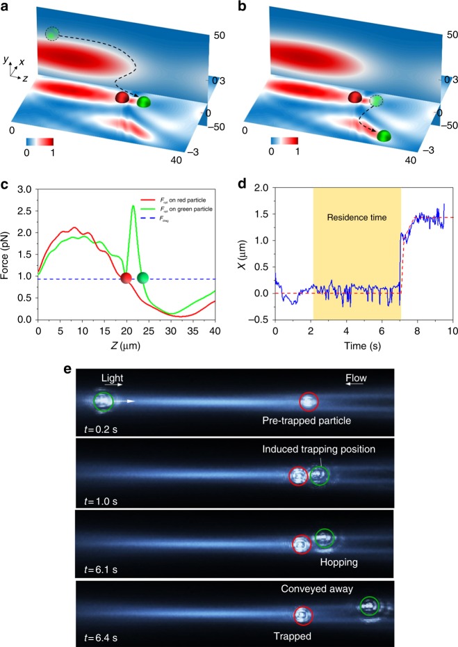
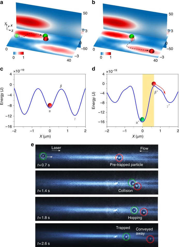
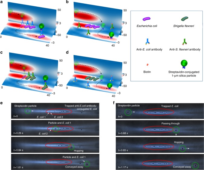
Particle trapping and binding in optical potential wells provide a versatile platform for various biomedical applications. However, implementation systems to study multi-particle contact interactions in an optical lattice remain rare. By configuring an optofluidic lattice, we demonstrate the precise control of particle interactions and functions such as controlling aggregation and multi-hopping. The mean residence time of a single particle is found considerably reduced from 7 s, as predicted by Kramer's theory, to 0.6 s, owing to the mechanical interactions among aggregated particles. The optofluidic lattice also enables single-bacteria-level screening of biological binding agents such as antibodies through particle-enabled bacteria hopping. The binding efficiency of antibodies could be determined directly, selectively, quantitatively and efficiently. This work enriches the fundamental mechanisms of particle kinetics and offers new possibilities for probing and utilising unprecedented biomolecule interactions at single-bacteria level.


Conflict of interest statement

The authors declare no competing financial interests.

Figures

Fig. 1
Fig. 1
The 2D controllable particle hopping in an optofluidic lattice. a Generation of the 2D lattice in an optofluidic chip. b Forces acting on a particle in the hotspot. c Illustration of the realisation of controllable particle hopping loop around the ellipsoid hotspots. Illustration of the simulated normalised light intensity (top row) and potential energy (bottom row) profiles of particle hopping triggered by three different mechanisms: d particle bypassing, e snookering-like particle collision and f particle aggregation. The intensity profiles in yz plane (vertical) are slightly shifted from the central line (x = 0) to have a better view of the particle trajectories
Fig. 2
Fig. 2
Particle hopping from the extra trapping position caused by particle pre-trapping. Illustration of a the green particle being trapped in the extra trapping position and b hopping to the adjacent potential well. Optical intensity profiles at the x–z plane (y = 0) and yz plane (x = 0) are plotted. c Force analysis on the 1 μm polystyrene particle along the z-direction when x = 0, y = 0. Blue line represents the drag force. Red and green lines represent the optical forces on red and green particles, respectively. d Projection of the green particle trajectory on the x-axis. e Experimental demonstration of the 1 μm particle hopping induced by the pre-trapped particle
Fig. 3
Fig. 3
Snookering-like particle collision-induced hopping between potential wells. Illustration of the particle a collision and b hopping combining with optical intensity profiles of the lattice. The energy profile of the potential wells c before and d after the particle collision, plotted along x-axis at y = 0 and z = 20 μm. e Experimental demonstration of the particle collision-induced hopping between potential wells
Fig. 4
Fig. 4
Particle hopping induced by particle aggregation. a Model for calculating mean first passage time of the particle in the potential well. b Illustration of hopping of the particle pair between adjacent hotspots. c Potential wells of total force Ft plotted along x-axis at y = 0 and z = 20 μm with different contact angles. d Calculated mean first passage time of the red particle varies with contact angle θ. Experimental residence time is distributed in the yellow area. e Experimental demonstration of the particle hopping induced by particle aggregation in the optofluidic lattice
Fig. 5
Fig. 5
Antibody selection through specific binding and hopping. E. coli and S. flexneri are trapped in the hotspot after they are conjugated with biotin-labelled anti-E. coli antibody and biotin-labelled anti-S. flexneri antibody, respectively. When a streptavidin-conjugated microparticle passes through the trapped a E. coli and c S. flexneri stained with specific antibodies, the microparticle is anchored to the bacterium through the very strong biotin–streptavidin interaction. The microparticle and bacterium hop together to another hotspot. When b E. coli and d S. flexneri are trapped in the hotspot after they are incubated with non-specific antibodies, the streptavidin-conjugated microparticle hops to another hotspot without binding with any bacterium due to the unsuccessful labelling of biotin. Experimental observation of the streptavidin-coated microparticle hopped with e E. coli conjugated with anti-E. coli antibody, and f passed away from bare E. coli due to unsuccessful conjugation of non-specific antibody (anti-S. flexneri antibody)

References

    1. Heller I, et al. STED nanoscopy combined with optical tweezers reveals protein dynamics on densely covered DNA. Nat. Methods. 2013;10:910–916. doi: 10.1038/nmeth.2599. - DOI - PubMed
    1. Perkins TT, Quake SR, Smith DE, Chu S. Relaxation of a single DNA molecule observed by optical microscopy. Science. 1994;264:822–826. doi: 10.1126/science.8171336. - DOI - PubMed
    1. Liao GB, Bareil PB, Sheng Y, Chiou A. One-dimensional jumping optical tweezers for optical stretching of bi-concave human red blood cells. Opt. Express. 2008;16:1996–2004. doi: 10.1364/OE.16.001996. - DOI - PubMed
    1. Sali A, Shakhnovich E, Karplus M. How does a protein fold? Nature. 1994;369:248–251. doi: 10.1038/369248a0. - DOI - PubMed
    1. White SH, Wimley WC. Membrane protein folding and stability: Physical principles. Annu. Rev. Biophys. Biomol. Struct. 1999;28:319–365. doi: 10.1146/annurev.biophys.28.1.319. - DOI - PubMed

Publication types

LinkOut - more resources