. 2018 Feb 26;9(1):842.
doi: 10.1038/s41467-018-03255-3.

Self-interaction of NPM1 modulates multiple mechanisms of liquid-liquid phase separation

Affiliations

Self-interaction of NPM1 modulates multiple mechanisms of liquid-liquid phase separation

Diana M Mitrea et al. Nat Commun. .

Abstract

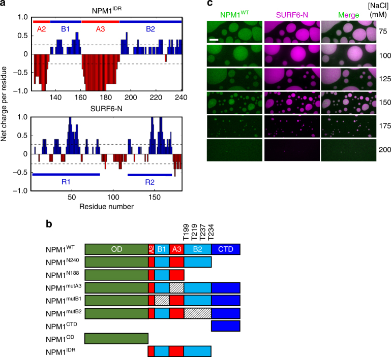
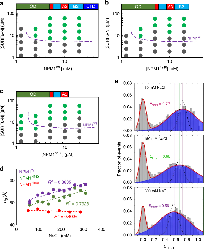
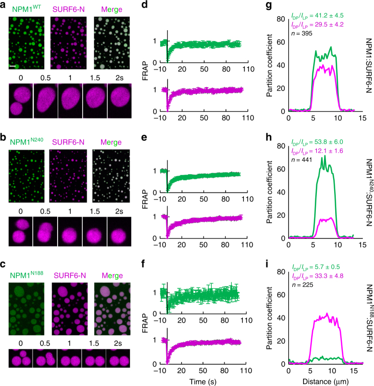
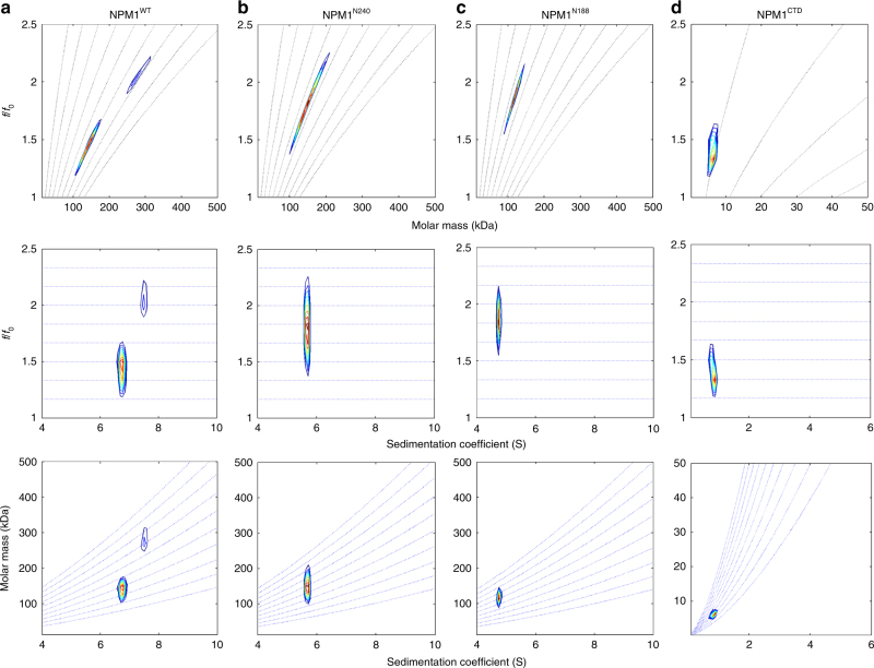
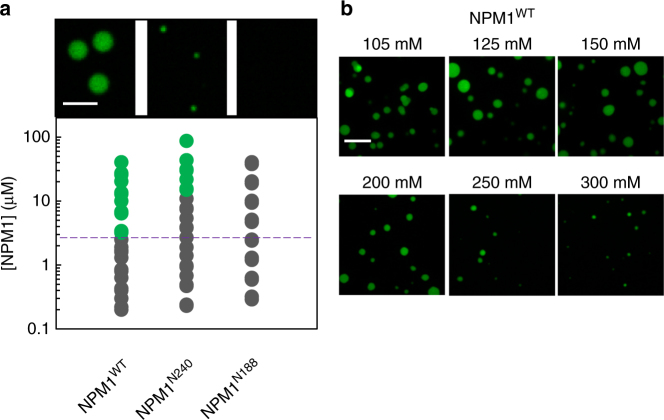
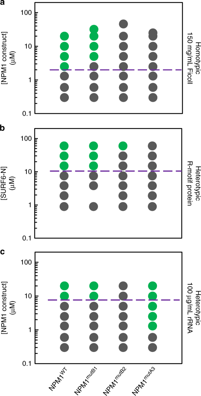
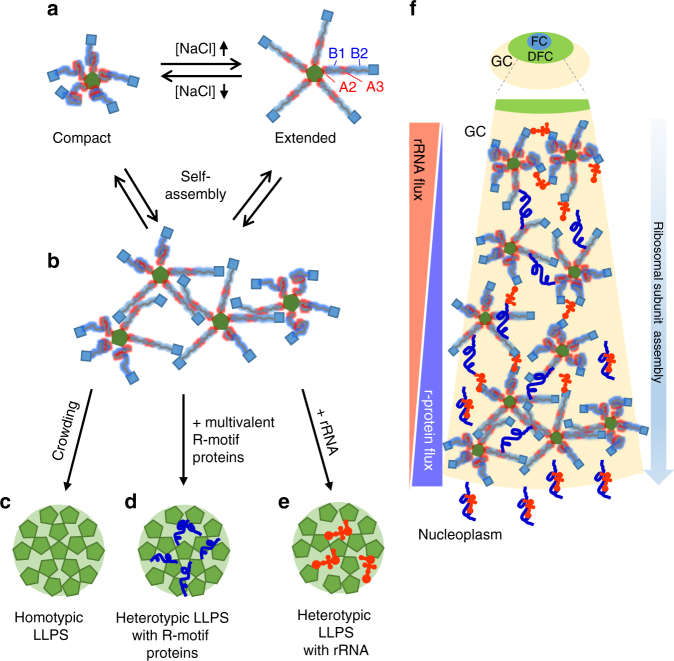
Nucleophosmin (NPM1) is an abundant, oligomeric protein in the granular component of the nucleolus with roles in ribosome biogenesis. Pentameric NPM1 undergoes liquid-liquid phase separation (LLPS) via heterotypic interactions with nucleolar components, including ribosomal RNA (rRNA) and proteins which display multivalent arginine-rich linear motifs (R-motifs), and is integral to the liquid-like nucleolar matrix. Here we show that NPM1 can also undergo LLPS via homotypic interactions between its polyampholytic intrinsically disordered regions, a mechanism that opposes LLPS via heterotypic interactions. Using a combination of biophysical techniques, including confocal microscopy, SAXS, analytical ultracentrifugation, and single-molecule fluorescence, we describe how conformational changes within NPM1 control valency and switching between the different LLPS mechanisms. We propose that this newly discovered interplay between multiple LLPS mechanisms may influence the direction of vectorial pre-ribosomal particle assembly within, and exit from the nucleolus as part of the ribosome biogenesis process.


Conflict of interest statement

The authors declare no competing financial interests.

Figures

Fig. 1
Fig. 1
Electrostatic interactions drive NPM1:SURF6-N LLPS. a Net charge per residue distribution using CIDER (http://pappulab.wustl.edu/CIDER/analysis/); top: the central intrinsically disordered region (IDR) in NPM1 exhibits polyampholityc properties; IDR contains two strong acidic tracts (A2 & A3) interleaved with moderately charged basic tracts (B1 & B2); bottom: the intrinsically disordered nucleolar protein SURF6 contains multivalent R-motifs throughout its primary structure. Data are shown for residues 1–182, corresponding to the construct used in this study (SURF6-N); b Schematic representation of the NPM1 constructs used in this study; c Confocal microscopy images of phase separation by 20 µM NPM1WT with 20 µM SURF6-N in buffer with variable concentrations of NaCl, as indicated. Screening of electrostatic interactions at high [NaCl] disrupts LLPS droplets. NPM1 is labeled with AlexaFluor 488 and SURF6-N with AlexaFluor 647. Scale bar, 10 µm
Fig. 2
Fig. 2
Intra-IDR interactions drive structural compaction within NPM1 and influence the threshold for heterotypic LLPS in the presence of SURF6-N. Phase diagrams determined by turbidity for NPM1WT (a), NPM1N240 (b), and NPM1N188 (c). The dotted purple line is a visual aid that represents the phase boundary for NPM1 between the mixed (gray circles) and demixed (green circles) states. (d) Changes in radii of gyration as a function of ionic strength, as determined from P(r) analysis of SAXS scattering curves; fitting errors are shown. (e) smFRET histograms showing the variation of NPM1 conformation at increasing [NaCl]. The solid lines represent fitting of the experimental data with a Gaussian model. The peak at zero is due to molecules lacking an active acceptor dye. The dotted line indicates the shot-noise simulation at each condition
Fig. 3
Fig. 3
NPM1 truncation mutants form liquid-like droplets in the presence of SURF6-N of differential composition. Fluorescence confocal microscopy images (ac) and FRAP curves (df) for droplets formed with 20 µM NPM1WT (a, d), NPM1N240 (b, e), and NPM1N188 (c, f) plus 20 µM SURF6-N. Representative curves illustrating cross-sections through droplets (gi) illustrating differences in the partition coefficients for the NPM1 constructs and SURF6-N; values represent mean ± s.d.; n = 5 or as indicated on graph
Fig. 4
Fig. 4
Two-dimensional size-and-shape distribution analyses of the sedimentation velocity AUC data. a NPM1, b NPM1N240, c NPM1N188, and d NPM1CTD. The transformed velocity data are shown as contour plots (heat maps) of c(M, f/f0,) (top), c(s, f/f0) (middle), and c(s, M) (bottom) with 0 fringes/S (white) to maximum value fringes/S (red), with increasing color temperature indicating higher values. Velocity data were acquired at 50,000 rpm at 20 °C in buffer comprised of 10 mM Tris, pH 7.5, 150 mM NaCl, 2 mM DTT. The sw,(f/f0)w, and M-values are listed in Supplementary Table 2
Fig. 5
Fig. 5
Only NPM1 constructs with an intact IDR undergo homotypic LLPS. a Phase diagrams for NPM1WT, NPM1N240 and NPM1N188 in buffer comprised of 10 mM Tris, 150 mM NaCl, 2 mM DTT, pH 7.5 in the presence of 150 mg/mL Ficoll PM70. The purple dotted line is a visual aid that represents the phase boundary for NPM1WT between the mixed (gray circles) and demixed (green circles) states. Representative microscopy images are shown for 20 µM protein samples. Scale bar = 5 µm; b confocal microscopy images of homotypic NPM1 droplets at 20 µM NPM1 in the presence of 150 mg/mL Ficoll PM70, in buffers containing the indicated NaCl concentration. Scale bar = 10 µm
Fig. 6
Fig. 6
Electrostatic interactions that drive conformational compaction allosterically couple the R-motif binding and rRNA binding modes of NPM1. Phase separation diagrams based on turbidity assays for the homotypic (a), heterotypic with SURF6-N (NPM1 constructs at 20 μM) (b) and heterotypic with rRNA (c) mechanisms, at the indicated NPM1 construct (a, c) or SURF6-N (b) concentrations. The purple dotted line is a visual aid that represents the phase boundary for NPM1WT between the mixed (gray circles) and demixed (green circles) states
Fig. 7
Fig. 7
NPM1 adopts a broad range of configurations to achieve biological multifunctionality. a Depending upon ionic conditions and available partners, NPM1 can adopt compact conformations, through intra-chain interactions between A- and B-tracts within its IDR and, extended conformations, and can assemble into multimers through in trans interactions between A- and B-tracts. b NPM1–NPM1 in trans interactions form the scaffold for homotypic LLPS. In a, b, and f, the NPM1 OD is represented as a green pentagon, the IDR as red and blue fuzzy lines representing the A- and B-tracts, respectively, and CTD as blue squares. Homotypic interactions (c) can be replaced by heterotypic interactions with multivalent R-motif proteins, such as SURF6 (curvy blue line) (d) or with rRNA (orange objects) (e), changing the nature of the scaffold. Note that, for visual clarity, the entire NPM1 pentamer is represented as a green pentagon in ce. f Schematic illustration of NPM1-mediated ribosomal subunit assembly in the GC of the nucleolus. Ribosomal subunits are comprised of rRNA and ribosomal proteins (r-proteins; curvy blue lines). rRNA is synthesized at the boundary between the fibrillar center (FC) and dense fibrillar component (DFC) and diffuses from the center to the periphery of the nucleolus, while the ribosomal proteins diffuse from the nucleoplasm into the nucleolus. In this model, the type of LLPS mechanism utilized by NPM1 within the GC depends upon the availability of its binding partners: rRNA, which moves outwards from the DFC/GC boundary, and R-motif proteins, which move inwards from the GC/nucleoplasm boundary, each drive different heterotypic LLPS mechanisms, in the central region of the GC. The close spatial proximity of rRNA and r-proteins within this central GC region, which exists through mixed heterotypic NPM1-mediated LLPS, enables the “hand-off” of these ribosomal components to interact with each other to form nascent ribosomal subunits (represented as conjoined orange objects and curvy blue lines). As ribosomal subunits assemble, their components no longer interact extensively with NPM1; at the same time, however, NPM1’s propensity for self-interaction compensates, maintaining the liquid-like GC scaffold through homotypic LLPS

References

    1. Mitrea DM, Kriwacki RW. Phase separation in biology; functional organization of a higher order. Cell Commun. Signal. 2016;14:1. doi: 10.1186/s12964-015-0125-7. - DOI - PMC - PubMed
    1. Banani SF, Lee HO, Hyman AA, Rosen MK. Biomolecular condensates: organizers of cellular biochemistry. Nat. Rev. Mol. Cell Biol. 2017;18:285–298. doi: 10.1038/nrm.2017.7. - DOI - PMC - PubMed
    1. Hyman AA, Weber CA, Julicher F. Liquid-liquid phase separation in biology. Annu. Rev. Cell Dev. Biol. 2014;30:39–58. doi: 10.1146/annurev-cellbio-100913-013325. - DOI - PubMed
    1. Banani SF, et al. Compositional control of phase-separated cellular bodies. Cell. 2016;166:651–663. doi: 10.1016/j.cell.2016.06.010. - DOI - PMC - PubMed
    1. Ahmad Y, Boisvert FM, Gregor P, Cobley A, Lamond AI. NOPdb: nucleolar proteome database--2008 update. Nucleic Acids Res. 2009;37:D181–D184. doi: 10.1093/nar/gkn804. - DOI - PMC - PubMed

Publication types

MeSH terms