. 2018 Feb 26;9(1):816.
doi: 10.1038/s41467-018-03105-2.

Hypoxia-inducible factor 2-alpha-dependent induction of amphiregulin dampens myocardial ischemia-reperfusion injury

Affiliations

Hypoxia-inducible factor 2-alpha-dependent induction of amphiregulin dampens myocardial ischemia-reperfusion injury

Michael Koeppen et al. Nat Commun. .

Abstract

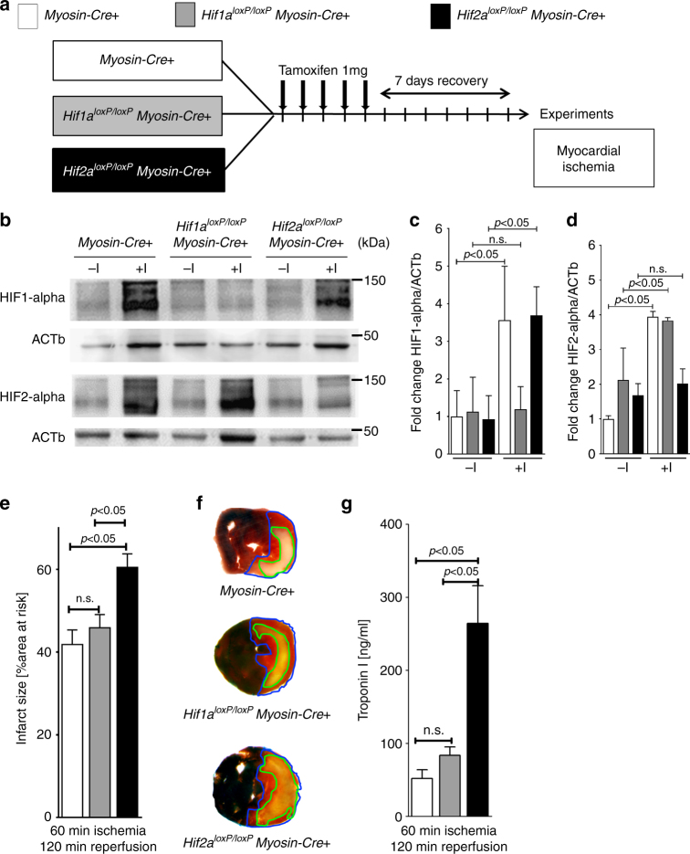
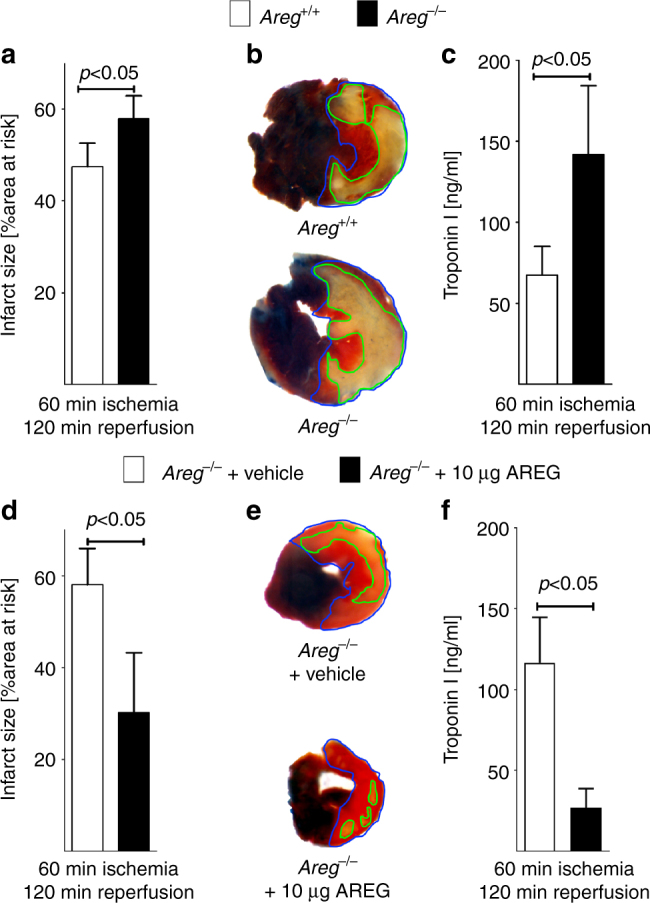
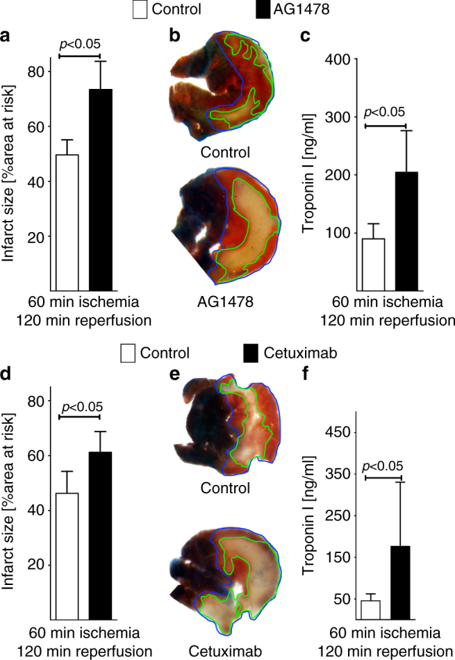
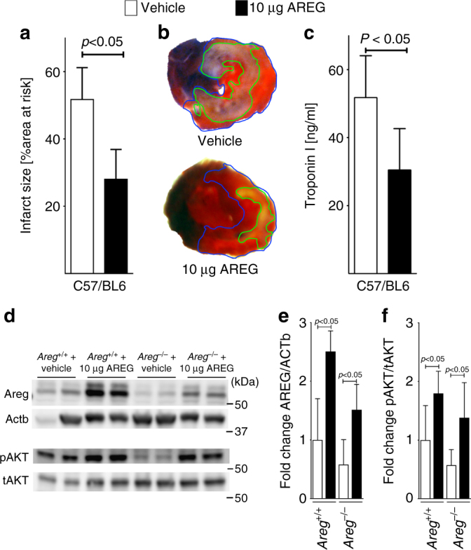
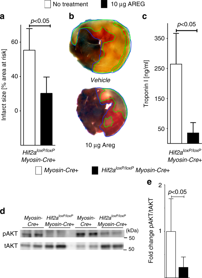
Myocardial ischemia-reperfusion injury (IRI) leads to the stabilization of the transcription factors hypoxia-inducible factor 1-alpha (HIF1-alpha) and hypoxia-inducible factor 2-alpha (HIF2-alpha). While previous studies implicate HIF1-alpha in cardioprotection, the role of HIF2-alpha remains elusive. Here we show that HIF2-alpha induces the epithelial growth factor amphiregulin (AREG) to elicit cardioprotection in myocardial IRI. Comparing mice with inducible deletion of Hif1a or Hif2a in cardiac myocytes, we show that loss of Hif2-alpha increases infarct sizes. Microarray studies in genetic models or cultured human cardiac myocytes implicate HIF2-alpha in the myocardial induction of AREG. Likewise, AREG increases in myocardial tissues from patients with ischemic heart disease. Areg deficiency increases myocardial IRI, as does pharmacologic inhibition of Areg signaling. In contrast, treatment with recombinant Areg provides cardioprotection and reconstitutes mice with Hif2a deletion. These studies indicate that HIF2-alpha induces myocardial AREG expression in cardiac myocytes, which increases myocardial ischemia tolerance.


Conflict of interest statement

The authors declare no competing financial interests.

Figures

Fig. 1
Fig. 1
Contribution of myocyte-specific hypoxia-inducible factor (HIF) isoforms Hif1a or Hif2a to cardioprotection. a Schematic of breeding approach to generate mice with induced myocyte-specific HIF deletions used in subsequent studies. Hif1aloxP/loxP or Hif2aloxP/loxP mice were crossed with Cre-recombinase expressing mice under the control of Myosin-heavy chain promoter (Myosin-Cre+); these mice express cre-recombinase under the control of a tamoxifen-inducer. Control animals (Myosin-Cre+), Hif1aloxP/loxPMyosin-Cre+ or Hif2aloxP/loxPMyosin-Cre+ received a daily dose of 1 mg i.p. Tamoxifen five consecutive days to induce the Cre-recombinase activity. After 7 days, animals underwent experimental protocol (60 min of in situ myocardial ischemia followed by 120 min of reperfusion). b HIF1-alpha or HIF2-alpha immunoblot analysis from homogenized myocardial tissue, harvested from male Myosin-Cre+ and Hif1aloxP/loxPMyosin-Cre+ or Hif2aloxP/loxPMyosin-Cre+ mice, matched in age and weight. Mice underwent a thoracotomy with no further treatment(-I) or 60 min of myocardial ischemia (+I) followed by 120 min reperfusion; β-Actin (ACTb) served as a loading control. One representative blot out of three experiments is shown. c, d Quantification by densitometry of the HIF-immunoblot results relative to ACTb. Data are expressed as mean fold change ±SD normalized to untreated myocardial tissue from Myosin-Cre+ compared by one-way ANOVA followed by Bonferroni’s multiple comparison test (n = 3 per group; c: F5,12 = 6.74, p = 0.0033; d: F5,12 = 21.85, p < 0.0001). e Infarct sizes ±SD in Myosin-Cre+, Hif1aloxP/loxP Myosin-Cre+ or Hif2aloxP/loxPMyosin-Cre+ mice, presented as the percentage to the area-at-risk after 60 min of ischemia, followed by 120 min of reperfusion (Myosin-Cre+ n = 5; Hif1aloxP/loxPMyosin-Cre+ n = 5; Hif2aloxP/loxPMyosin-Cre+ n = 4; per group mean ± SD; compared by one-way ANOVA followed by Bonferroni’s multiple comparison test; F2,11 = 7.901; p = 0.0075). f Representative infarct staining from Myosin-Cre+, Hif1aloxP/loxPMyosin-Cre+ or Hif2aloxP/loxPMyosin-Cre+. g Troponin serum levels after 60 min ischemia, followed by 120 min of reperfusion in Myosin-Cre+, Hif1aloxP/loxPMyosin-Cre+ or Hif2aloxP/loxPMyosin-Cre+ (Myosin-Cre+ n = 5; Hif1aloxP/loxPMyosin-Cre+ n = 5, and Hif2aloxP/loxPMyosin-Cre+ n = 4 per group; presented as mean ± SD; compared by one-way ANOVA followed by Bonferroni’s multiple comparison test; F2,11 = 19.14, p = 0.0003)
Fig. 2
Fig. 2
Hif2a-dependent induction of amphiregulin during myocardial injury. a, b Myosin-Cre+ or Hif2aloxP/loxPMyosin-Cre+ underwent 45 min myocardial ischemia, followed by 120 min reperfusion. After harvest of the area-at-risk, total RNA was probed for transcriptional changes using a microarray technique (Agilent-014868 Whole Mouse Genome Microarray 4x44K G4122F Probe Name version). Transcriptional changes were calculated in relation to the myocardial baseline expression in untreated littermate controls of the same genotype. Array data have been deposited at http://www.ncbi.nlm.nih.gov/geo/ (accession number GSE67308). a List of “top hits” of genes transcriptionally induced in the Area-At-Risk in Myosin-Cre+ but unchanged in Hif2aloxP/loxPMyosin-Cre+ (n = 4 per group, mean ± SD). Note: the most prominent differential regulation was found for the epidermal growth factor amphiregulin (Areg). b Analysis of relevant networks in the IPA (Ingenuity Pathway Analysis®) library most significant to the dataset. Note: selective induction of Areg c Myosin-Cre+, Hif1aloxP/loxPMyosin-Cre+, or Hif2aloxP/loxPMyosin-Cre+ underwent 60 min myocardial ischemia, following 120 min reperfusion. Total RNA from the area-at-risk was probed for transcriptional changes of Areg by real-time RT-PCR. Transcriptional changes were calculated relative to an internal housekeeping gene (Actb). Data are expressed as mean fold change ± SD compared to untreated myocardial tissue from littermates of the respective genotype compared by one-way ANOVA followed by Bonferroni’s multiple comparison test (Myosin-Cre+: −I n = 6, +I n = 5; Hif1aloxP/loxPMyosin-Cre+: −I n = 3; +I n = 4; Hif2aloxP/loxPMyosin-Cre+: −I n = 6; +I n = 7; F5,25 = 33.53; p < 0.0001). d AREG immunoblot performed for protein isolated from the ischemic myocardial tissue after 60 min of ischemia and 120 min reperfusion or untreated littermate controls of Myosin-Cre+, Hif1aloxP/loxPMyosin-Cre+ or Hif2aloxP/loxPMyosin-Cre+, respectively. A representative image of three individual experiments is presented. e Quantification by densitometry of the AREG immunoblot results relative to ACTb. Data are expressed as mean fold change ± SD normalized to untreated myocardial tissue from Myosin-Cre+ compared by one-way ANOVA followed by Bonferroni’s multiple comparison test (n = 3 per group; F5,12 = 4.487; p = 0.0154)
Fig. 3
Fig. 3
Functional role of HIF2A in the transcriptional regulation of amphiregulin (AREG) in human cardiac myocytes. a–e Human cardiac myocytes (HCM) underwent control, HIF1A-specific or HIF2a-specific short hairpin RNA (shRNA) lentiviral transfection to suppress transcription of hypoxia-inducible factors HIF1A or HIF2A. a, b shRNA-transfected HCM were exposed to ambient hypoxia (1% oxygen) for 16 h and analysis of transcript changes by RT-PCR of HIF1A or HIF2A, respectively. Transcriptional changes were calculated relative to an internal housekeeping gene (Actin-b). Data are expressed as mean fold change ± SD compared to normoxic cells (n = 6 per group). c Immunoblot for HIF1A or HIF2A from shRNA-transfected normoxic or hypoxic HCM. β-Actin (ACTb) served as a loading control. d, e Quantification by densitometry of the HIF1-alpha or HIF2-alpha immunoblot results relative to ACTb. Data are expressed as mean fold change ± SD normalized to shControl compared by one-way ANOVA followed by Bonferroni’s multiple comparison test (n = 3 per group; d F5,11 = 14.48, p = 0.0002; e F5,11 = 6,726, p = 0.0042). f HCM transfected with shRNA directed against HIF1A, HIF2A or control shRNA were exposed to ambient hypoxia (1% oxygen) for 16 h or were maintained under normoxic conditions (21% oxygen). Subsequently, total RNA was isolated and probed by RT-PCR for transcriptional changes of amphiregulin (AREG). Data are expressed as mean fold change ± SD compared to normoxic cells (n = 3 per group). Transcriptional changes were calculated relative to an internal housekeeping gene (ACTb). Data compared by one-way ANOVA followed by Bonferroni’s multiple comparison test (F5,12 = 14.33; p = 0.0001). g HCM transfected with shRNA directed against HIF1A, HIF2A, or control shRNA, total protein was isolated and immunoblotted for AREG. b-Actin (Actb) served as a loading control. One representative blot out of three experiments is shown. h Quantification by densitometry of the HIF immunoblot results relative to ACTb. Data are expressed as mean fold change ± SD normalized to normoxic cells (shControl) and compared by one-way ANOVA followed by Bonferroni’s multiple comparison test (n = 3 per group; F5,12 = 11, 58; p = 0.0003)
Fig. 4
Fig. 4
Cardiac amphiregulin protein levels in myocardial tissues from healthy controls or patients with ischemic heart disease (IHD). a Amphiregulin (AREG) protein levels were determined by immunoblot after total protein isolation from control, healthy myocardial tissue (labeled C), or samples from patients with ischemic heart disease (labeled IHD). β-Actin (ACTb) served as a loading control. b Quantification by densitometry of the AREG immunoblot results relative to ACTb (n = 10 in both groups; data compared by two-sided Mann–Whitney test)
Fig. 5
Fig. 5
Myocardial tissue injury in Areg−/− mice exposed to myocardial ischemia and reperfusion injury. af Experimental animals were exposed to 60 min of myocardial ischemia, followed by 120 min of reperfusion; infarct sizes were measured by double staining with Evan’s blue and triphenyltetrazolium chloride and serum samples were collected. All infarct sizes are presented as the percentage of infarcted tissue in relation to the area-at-risk. Serum troponin levels were determined by ELISA (a). Infarct sizes were determined in gene-targeted mice for amphiregulin (Areg−/− mice) or isogenic control animals (Areg+/+ mice) (n = 5 per group). b Representative infarct staining from Areg−/− or Areg+/+. c Troponin serum levels in Areg−/− or Areg+/+ mice (n = 5 per group). d Reconstitution of Areg−/− mice with recombinant AREG. Infarct sizes in Areg−/− treated with vehicle or 10 μg of recombinant murine Areg, administered over 15 min period by an indwelling catheter (n = 6 per group). e Representative infarct staining from Areg−/− mice treated with vehicle or 10 µg recombinant murine Areg. f Troponin serum levels after myocardial IR injury of Areg−/− mice treated with vehicle or 10 µg recombinant murine Areg via an intraarterial catheter over a 15 min period (n = 7 per group). All data presented as mean ± SD. Statistical significance assessed by two-sided, unpaired Student’s t-test
Fig. 6
Fig. 6
Pharmacologic inhibition of the amphiregulin receptor ErbB1 during murine myocardial ischemia–reperfusion injury. ac Mice underwent 60 min of myocardial ischemia, followed by 120 min of reperfusion; infarct sizes were measured by double staining with Evan’s blue and triphenyltetrazolium chloride and serum samples were collected. All infarct sizes are presented as the percentage of infarcted tissue in relation to the area-at-risk. Serum Troponin levels were determined by ELISA. a Infarct sizes of C57/BL6 mice after 60 min of ischemia and 120 min reperfusion, treated prior to myocardial ischemia reperfusion with vehicle or an ERBB1-specific inhibitor (AG1478), administered 15 min prior to ischemia via an indwelling arterial catheter. Data presented as the percentage of infarcted area in relation to area-at-risk (n = 4 per group). b Representative infarct staining of C57/BL6 treated with vehicle or AG1478. c Troponin serum levels of C57/BL6 mice, after 60 min ischemia, 120 min reperfusion, treated with vehicle or AG1478 (n = 5 per group). d Infarct sizes of C57/BL6 mice after 60 min of ischemia and 120 min reperfusion, treated 15 min prior to ischemia via an indwelling arterial catheter with vehicle or a highly specific ERBB1-specific inhibitor (Cetuximab). Data presented as the percentage of infarcted area in relation to area-at-risk (control n = 4; cetuximab n = 8). e Representative infarct staining of C57/BL6 treated with vehicle or Cetuximab. f Troponin serum levels of C57/BL6 mice after 60 min ischemia, 120 min reperfusion, treated with vehicle or Cetuximab (n = 7 per group). All data in this figure is presented as mean ± SD. Statistical significance assessed by two-sided, unpaired Student’s t-test
Fig. 7
Fig. 7
Treatment of murine myocardial ischemia and reperfusion injury with recombinant amphiregulin. ac Wild-type were exposed to 60 min of myocardial ischemia, followed by 120 min of reperfusion; infarct sizes were measured by double staining with Evan’s blue and triphenyltetrazolium chloride and serum samples were collected. All Infarct sizes are presented as the percentage of infarcted tissue in relation to the area-at-risk. Serum troponin levels were determined by ELISA. a Infarct sizes of C57/BL6 mice after 60 min of ischemia and 120 min reperfusion, treated with vehicle or 10 µg recombinant murine AREG, administered over a 15 min period via an indwelling arterial catheter. Data presented are the percentage of infarcted area in relation to area-at-risk. Statistical significance assessed by two-sided, unpaired Student’s t-test (n = 7 per group; data presented as mean ± SD). b Representative infarct staining of C57/BL6 treated with vehicle or 10 µg recombinant murine AREG. c Serum troponin levels of C57/BL6 mice, after 60 min ischemia, 120 min reperfusion, treated with vehicle or 10 µg recombinant murine Areg. Statistical significance assessed by two-sided, unpaired Student’s t-test (n = 5 per group; data presented as mean ± SD). d Areg+/+ or Areg−/− mice received treatment with vehicle or 10 μg of recombinant murine AREG, administered over a 15 min period by an indwelling catheter. The animals underwent 60 min ischemia and 120 min reperfusion, followed by total protein isolation form the area-at-risk. Upper panel: protein was immunoblotted for AREG, β-actin (ACTb) served as a loading control. Lower panel: protein was immunoblotted for total AKT (tAKT) and phosphorylated AKT (pAKT), respectively. Two samples for each condition and genotype are presented. One representative blot out of three experiments is shown. e, f Quantification by densitometry of the AREG and pAKT immunoblot results relative to ACTb or Total AKT (tAKT). Data are expressed as mean fold change ± SD normalized to vehicle treated Areg+/+ and compared by one-way ANOVA followed by Bonferroni’s multiple comparison test (Areg+/++Vehicle n = 5; all other groups n = 6 mice per group, e F3,19 = 23,11, p < 0.0001; f F3,20 = 9,549, p = 0.0002)
Fig. 8
Fig. 8
Reconstitution of cardiomyocyte-specific Hif2a-deficient mice with recombinant amphiregulin. ac Hif2aloxP/loxPMyosin-Cre+ of similar age, gender, and weight as control mice were exposed to 60 min of myocardial ischemia, followed by 120 min of reperfusion; infarct sizes were measured by double staining with Evan’s blue and triphenyltetrazolium chloride and serum samples were collected. All Infarct sizes are presented as the percentage of infarcted tissue in relation to the area-at-risk. Serum troponin levels were determined by ELISA. Note that data in (ac) in the “no treatment group” are used in part in Fig. 1c–e to display and analyze IR injury from similar experimental conditions. a Infarct sizes of Hif2aloxP/loxPMyosin-Cre+ mice after 60 min of ischemia and 120 min reperfusion that were pre-treated with 10 µg recombinant murine AREG administered over a 15 min period via an indwelling arterial catheter or received no pharmacologic intervention. Data presented as the percentage of infarcted area in relation to area-at-risk. Statistical significance assessed by two-sided, unpaired Student’s t-test (no treatment n = 6; 10 μg AREG n = 9, data presented as mean ± SD). b Representative infarct staining of Hif2aloxP/loxPMyosin-Cre+ that were pre-treated with 10 µg recombinant murine Areg administered over a 15 min period via an indwelling arterial catheter or received no pharmacologic intervention. c Serum troponin levels of Hif2aloxP/loxPMyosin-Cre+ mice, after 60 min ischemia, 120 min reperfusion that were pre-treated with 10 µg recombinant murine AREG administered over a 15 min period via an indwelling arterial catheter, or received no pharmacologic intervention. Statistical significance assessed by two-sided, unpaired Student’s t-test (no treatment n = 4; 10 μg AREG n = 7, data presented as mean ± SD). d Hif2aloxP/loxPMyosin-Cre+ or Myosin-Cre+ mice underwent 60 min myocardial ischemia, followed by 120 min of reperfusion. Then the area-at-risk was excised, protein isolated and immunoblotted for total AKT (tAKT) or phosphorylated Akt (pAKT). Two samples for each condition and genotype are presented. One representative blot out of three experiments is shown. e Quantification by densitometry of pAKT immunoblot results relative to total AKT (tAKT). Data are expressed as mean fold change ± SD normalized to Myosin-Cre+. Statistical significance assessed by two-sided, unpaired Student’s t-test (three individual blots analyzed with n = 7 mice per group)

References

    1. Eltzschig HK, Eckle T. Ischemia and reperfusion—from mechanism to translation. Nat. Med. 2011;17:1391–1401. doi: 10.1038/nm.2507. - DOI - PMC - PubMed
    1. Eltzschig HK, Bratton DL, Colgan SP. Targeting hypoxia signalling for the treatment of ischaemic and inflammatory diseases. Nat. Rev. Drug Discov. 2014;13:852–869. doi: 10.1038/nrd4422. - DOI - PMC - PubMed
    1. Yellon DM, Hausenloy DJ. Myocardial reperfusion injury. N. Engl. J. Med. 2007;357:1121–1135. doi: 10.1056/NEJMra071667. - DOI - PubMed
    1. Eltzschig HK, Bonney SK, Eckle T. Attenuating myocardial ischemia by targeting A2B adenosine receptors. Trends Mol. Med. 2013;19:345–354. doi: 10.1016/j.molmed.2013.02.005. - DOI - PMC - PubMed
    1. Botker HE, et al. Remote ischaemic conditioning before hospital admission, as a complement to angioplasty, and effect on myocardial salvage in patients with acute myocardial infarction: a randomised trial. Lancet. 2010;375:727–734. doi: 10.1016/S0140-6736(09)62001-8. - DOI - PubMed

Publication types

MeSH terms