. 2018 Feb 26;8(1):3603.
doi: 10.1038/s41598-018-22039-9.

HIF1α-dependent glycolysis promotes macrophage functional activities in protecting against bacterial and fungal infection

Affiliations

HIF1α-dependent glycolysis promotes macrophage functional activities in protecting against bacterial and fungal infection

Chunxiao Li et al. Sci Rep. .

Abstract

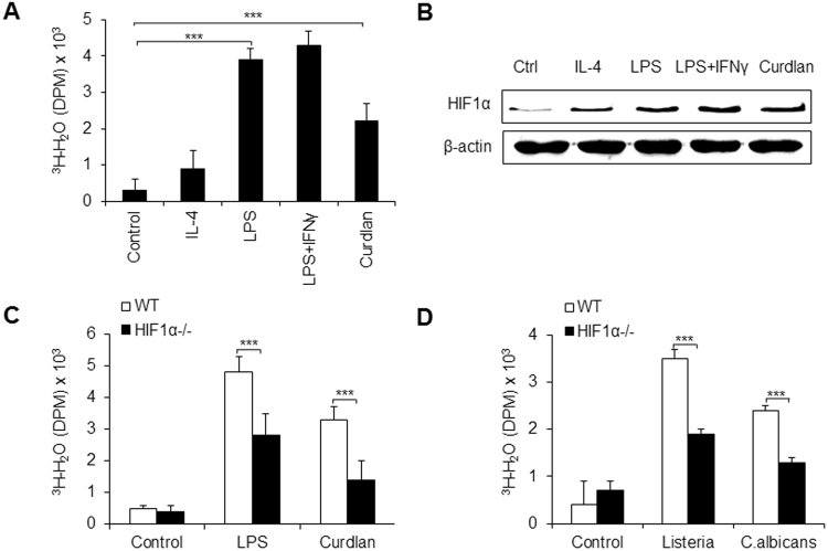
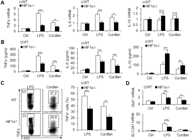
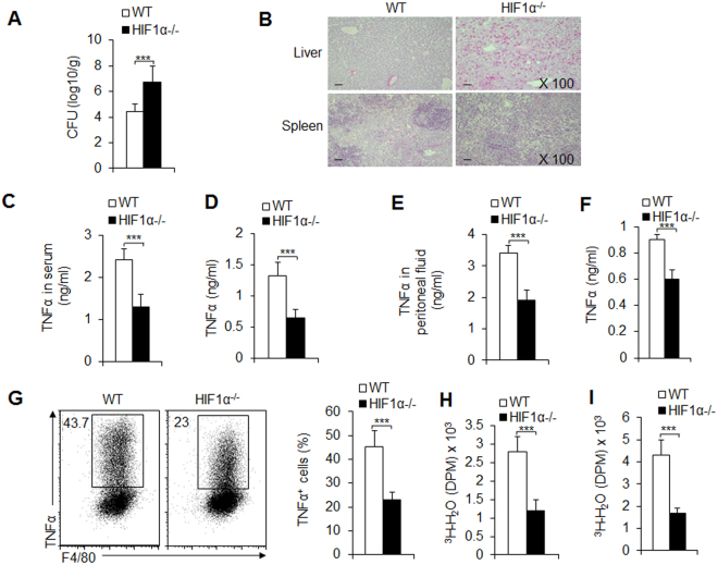
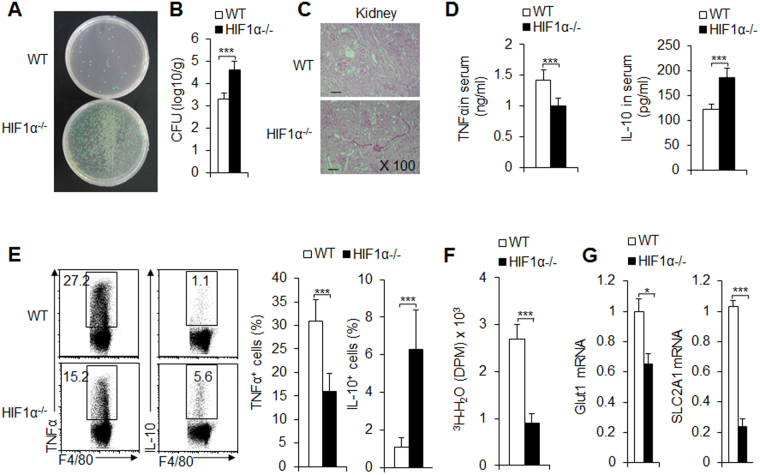
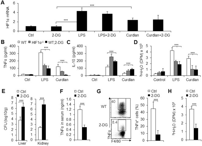
Macrophages are important innate immune defense system cells in the fight against bacterial and fungal pathogenic infections. They exhibit significant plasticity, particularly with their ability to undergo functional differentiation. Additionally, HIF1α is critically involved in the functional differentiation of macrophages during inflammation. However, the role of macrophage HIF1α in protecting against different pathogenic infections remains unclear. In this study, we investigated and compared the roles of HIF1α in different macrophage functional effects of bacterial and fungal infections in vitro and in vivo. We found that bacterial and fungal infections produced similar effects on macrophage functional differentiation. HIF1α deficiency inhibited pro-inflammatory macrophage functional activities when cells were stimulated with LPS or curdlan in vitro or when mice were infected with L. monocytogenes or C. albicans in vivo, thus decreasing pro-inflammatory TNFα and IL-6 secretion associated with pathogenic microorganism survival. Alteration of glycolytic pathway activation was required for the functional differentiation of pro-inflammatory macrophages in protecting against bacterial and fungal infections. Thus, the HIF1α-dependent glycolytic pathway is essential for pro-inflammatory macrophage functional differentiation in protecting against bacterial and fungal infections.


Conflict of interest statement

The authors declare no competing interests.

Figures

Figure 1
Figure 1
HIF1α-dependent glycolysis is associated with pro-inflammatory macrophage differentiation during inflammation. Sorted peritoneal exudate macrophages (PEMs) from C57BL/6 mice were stimulated with IL-4 (1000 U), LPS (100 ng/mL), LPS + IFNγ (100 ng/mL) or curdlan (100 ng/mL) for 10–12 h, and the glycolytic pathway activity was determined by the generation of 3H-labeled H2O from [3-3H]-glucose (A). HIF1α protein expression was determined using immunoblot (B). Sorted PEMs from wild-type (WT) C57BL/6 mice or Hif1αflox/flox; Lyz-Cre (HIF1α−/−) mice were stimulated with the indicated stimuli for 10–12 h, and the glycolytic pathway activity was determined (C,D). Data is presented as the means ± SD (n = 4). One representative experiment of three to four independent experiments is shown. *P < 0.05 and ***P < 0.001, compared with the indicated groups.
Figure 2
Figure 2
HIF1α is critical for pro-inflammatory macrophage differentiation in vitro. Sorted PEMs from WT and HIF1α−/− mice were stimulated with LPS (100 ng/mL) or curdlan (100 ng/mL) for 10–12 h, and the indicated mRNA expression was determined with qPCR (A). Supernatant was collected and the concentration of the indicated cytokines was determined with an ELISA (B). (C) Peritoneal exudate cells from WT and HIF1α−/− mice were activated with the indicated stimuli, and the intracellular expression of TNFα in F4/80+ macrophages was determined with flow cytometry; a representative image is shown in the left figure, and data are summarized in the right figure. (D) Sorted PEMs were stimulated with LPS or curdlan for 10–12 h, and the mRNA expression of glycolytic pathway associated molecules was determined with qPCR. Data are presented as the means ± SD (n = 3–5). One representative experiment of three independent experiments is shown. *P < 0.05; **P < 0.01 and ***P < 0.001, compared with the indicated groups; n.s. = not significant.
Figure 3
Figure 3
HIF1α is required for pro-inflammatory macrophage differentiation following Listeria bacterial infection. C57BL/6 WT or HIF1α−/− mice were i.v. injected with 1 × 105 CFU of L. monocytogenes bacteria. 48 h after infection, mouse livers were collected, and the CFU were determined (A). Infected mice developed severe infection and inflammatory cell infiltration, as shown by the histological staining of H&E (B). At the same time point after infection, serum or peritoneal exudate TNFα levels were determined using an ELISA (C and E); the sorted splenic macrophages or PEMs were stimulated with LPS for 12 h, the supernatant was collected, and TNFα levels were determined using an ELISA (D and F); the glycolytic pathway activity of macrophages was also determined (H and I). TNFα expression in F4/80+ macrophages from peritoneal exudates was analyzed with FCM. A representative figure is shown in the left image, and the data are summarized in the right image (G). Data are presented as the means ± SD (n = 3–5). One representative experiment of three independent experiments is shown. ***P < 0.001, compared with the indicated groups.
Figure 4
Figure 4
HIF1α is required for pro-inflammatory macrophage differentiation following C. albicans fungal infection. C57BL/6 WT or HIF1α−/− mice were i.v. injected with 2 × 105 of live C. albicans yeast. After 9 days, mice were killed for analysis. Infected kidneys were collected and the fungal burden in the kidneys is shown (A), expressed as CFU per g (B), and an image of the H&E staining of pathological kidney injuries is shown (C); serum was collected, and the indicated cytokine was determined using an ELISA (D). At the same time point during infection, the PEMs were collected, and the indicated cytokine expression was analyzed with FCM; a representative figure is shown in the left image, and data are summarized in the right image (E). The glycolytic pathway activity of PEMs was also determined (F,G). Data are presented as the means ± SD (n = 3–5). One representative experiment of three independent experiments is shown. *P < 0.05 and ***P < 0.001, compared with the indicated groups.
Figure 5
Figure 5
Glycolytic pathway activity was required for pro-inflammatory macrophage differentiation. PEMs with indicated treatments for 10–12 h (2-DG, 1 mmol/L; LPS, 100 ng/mL; curdlan, 100 ng/mL; AD). A. HIF1α mRNA expression of PEMs was determined with qPCR (Value of control groups was set to 1). (B and C) Supernatants were collected, and the indicated cytokine concentration was determined using an ELISA. (D) The glycolytic pathway activity was summarized. Mice were i.p. injected with 1 × 105 of C. albicans yeast and also injected intraperitoneally with 2-DG (2 g/kg body weight) or solvent alone (PBS; Ctrl) for 9 days. 2-DG or PBS were given daily up until the day before the mice were euthanized (EH). (E) Mouse liver and kidneys were collected, and the CFU was evaluated. (F) Serum TNFα concentration (F). (G) TNFα expression of PEMs with FCM. A representative image is shown in the left figure and data are summarized in the right figure. (H) The glycolytic pathway activity of PEMs. Data are presented as the means ± SD (n = 3–4). One representative experiment of three independent experiments is shown. P < 0.001, compared with the indicated groups.
Figure 6
Figure 6
Pharmacologically targeting HIF1α and glycolytic pathway activity in mouse and human cells. Mouse PEMs (AE) or human macrophages (FJ) pulsed with C. albicans yeast (1 × 105) for 3 days in the absence or presence of CoCl2 (200 µM), 2-ME (2 µM), or 2-DG (1 mmol/L). (A and E) The HIF1α mRNA expression of macrophages was determined with qPCR. (B and F) The TNFα concentration of supernatants was determined. (C and G) The CFU was evaluated. (D and H) The Glut1 mRNA expression was determined with qPCR. Data are presented as the means ± SD (n = 3–4). One representative experiment of three independent experiments is shown. *P < 0.05 and ***P < 0.001, compared with the indicated groups.

References

    1. Murray PJ, et al. Macrophage activation and polarization: nomenclature and experimental guidelines. Immunity. 2014;41:14–20. doi: 10.1016/j.immuni.2014.06.008. - DOI - PMC - PubMed
    1. Murray, P. J. Macrophage Polarization. Annual review of physiology (2016). - PubMed
    1. Izquierdo E, et al. Reshaping of Human Macrophage Polarization through Modulation of Glucose Catabolic Pathways. J Immunol. 2015;195:2442–2451. doi: 10.4049/jimmunol.1403045. - DOI - PubMed
    1. Liu G, Yang H. Modulation of macrophage activation and programming in immunity. Journal of cellular physiology. 2013;228:502–512. doi: 10.1002/jcp.24157. - DOI - PubMed
    1. Jha AK, et al. Network integration of parallel metabolic and transcriptional data reveals metabolic modules that regulate macrophage polarization. Immunity. 2015;42:419–430. doi: 10.1016/j.immuni.2015.02.005. - DOI - PubMed

Publication types

Substances