. 2018 Mar;25(3):252-260.
doi: 10.1038/s41594-018-0037-5. Epub 2018 Feb 26.

Cryo-EM and X-ray structures of TRPV4 reveal insight into ion permeation and gating mechanisms

Affiliations

Cryo-EM and X-ray structures of TRPV4 reveal insight into ion permeation and gating mechanisms

Zengqin Deng et al. Nat Struct Mol Biol. 2018 Mar.

Abstract

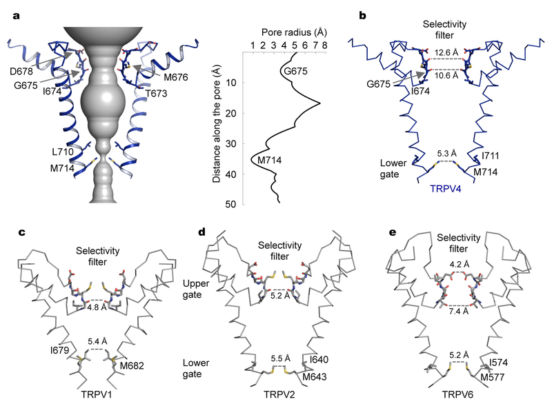
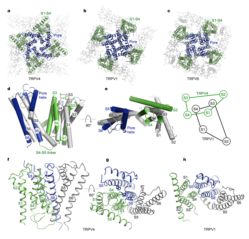
The transient receptor potential (TRP) channel TRPV4 participates in multiple biological processes, and numerous TRPV4 mutations underlie several distinct and devastating diseases. Here we present the cryo-EM structure of Xenopus tropicalis TRPV4 at 3.8-Å resolution. The ion-conduction pore contains an intracellular gate formed by the inner helices, but lacks any extracellular gate in the selectivity filter, as observed in other TRPV channels. Anomalous X-ray diffraction analyses identify a single ion-binding site in the selectivity filter, thus explaining TRPV4 nonselectivity. Structural comparisons with other TRP channels and distantly related voltage-gated cation channels reveal an unprecedented, unique packing interface between the voltage-sensor-like domain and the pore domain, suggesting distinct gating mechanisms. Moreover, our structure begins to provide mechanistic insights to the large set of pathogenic mutations, offering potential opportunities for drug development.


Conflict of interest statement

Competing Financial Interests

The authors declare no competing financial interests.

Figures

Fig. 1 |
Fig. 1 |. A TRPV4 construct for structural analyses.
a, Domain organization of the Xenopus tropicalis TRPV4 channel. The crystallization and cryo-EM construct comprises residues 133–797 with mutation N647Q eliminating glycosylation. b, 86Rb+ flux in transfected CosM6 cells. Activation of the full-length wild type channel (black squares, n = 3, mean ± SEM) and the truncated construct TRPV4cryst (gray squares, n = 3, mean ± SEM) by 1 μM GSK101 is evaluated by relative efflux of 86Rb+, in comparison with cells transfected with an empty vector (empty circles and squares, n = 3, mean ± SEM). Squares and circles indicate measurements with and without GSK101, respectively. cd, TRPV4 (c) and TRPV4cryst (d) currents in an inside-out membrane patch upon application of GSK101 (50 nM in C and 5 μM in D) from the cytoplasmic side. Unitary openings of ~ 8 pA at −30 mV are shown on the middle panels. The two first channel opening events are labeled as O1 and O2. All point histograms at the lower panels represent 5 seconds of traces. Wild type Xenopus TRPV4 has a unitary conductance of 252 ± 22 pS at −30 mV membrane in symmetric 150 mM KCl, while TRPV4cryst under the same conditions has a conductance of 264 ± 19 pS.
Fig. 2 |
Fig. 2 |. Cryo-EM structure of TRPV4.
a, Cryo-EM reconstruction of the tetrameric channel. Each subunit is uniquely colored, and the membrane boundary is indicated as gray lines. b, Orthogonal views of the overall structure. Subunits are colored as in (a). c, Structure of a single subunit. Each domain is labeled and uniquely colored.
Fig. 3 |
Fig. 3 |. Ion permeation pathway.
a, The ion conduction pore of TRPV4, shown as gray surface calculated with HOLE. Only two opposing subunits are shown for clarity. A single constriction formed by M714 residues defines the intracellular gate in S6. The pore radius along the permeation pathway is shown in the right panel. b, Details of the TRPV4 pore. Comparison with other TRPV pore structures shown in (ce) reveals a closed lower gate and the absence of an upper gate in TRPV4. ce, The pore structures of TRPV1 (PDB: 3J5P, c), TRPV2 (PDB: 5AN8, d), and TRPV6 (PDB: 5IWK, e), showing two constrictions: an upper gate in the selectivity filter and a lower gate in S6.
Fig. 4 |
Fig. 4 |. Ion binding in the pore.
ac, Side views of the TRPV4 pore with a bound Cs+ (magenta, a), Ba2+ (Orange, b), and Gd3+ (Cyan, c) respectively. Only two opposing subunits are shown for clarity. Ions are shown as spheres. The anomalous difference electron densities for Cs+ (magenta, a), Ba2+ (Orange, b), and Gd3+ (Cyan, c) are shown as meshes, contoured at 5.0 σ, 7.0 σ, and 6.0 σ respectively. d, Top view of the pore with a bound Gd3+. All four subunits are shown and the contour level is the same as in (c). e, Top view of the pore showing strong densities potentially corresponding to tightly bound lipid molecules. f, Side view of the densities as in (e).
Fig. 5 |
Fig. 5 |. Structural comparisons of the transmembrane domains of TRPV channels.
ac, Structures of TRPV4 (a), TRPV1 (b) (PDB: 3J5P), and TRPV6 (c) (PDB: 5IWK). The S1–S4 domains and the pore domains are highlighted in green and blue respectively. d and e, Orthogonal views of TRPV4 (green and blue) and TRPV1 (gray) transmembrane domains aligned by their pore domains (all four S5 and S6 helices). Also shown in (e) is a schematic diagram illustrating the re-orientation of the S1–S4 domain in TRPV4. f and g, Two views showing extensive packing interactions between helices S3 and S4 from one subunit and helices S5 and S6 from an adjacent subunit in TRPV4. Side chains of tightly packed residues are shown as sticks. h, A similar view as in (g) for TRPV1, showing that only S4 makes considerable contacts with the pore domain from an adjacent subunit.
Fig. 6 |
Fig. 6 |. A map for TRPV4 channelopathies.
a, Disease mutations are mapped onto the structure of a single subunit (gray ribbon). Mutations causing skeletal dysplasias (SD), peripheral neuropathy, and osteoarthropathy are shown as red, blue, and green spheres, respectively. For consistency, labeled residues are numbered according to the human TRPV4 sequence. b, Osteoarthropathy mutations in finger 3. F273 (shown as green sticks) interacts with a network of aromatic residues (shown as sticks) both within the same subunit and from the β sheet (shown in cyan) of an adjacent subunit. c, SD mutations in the S4–S5 linker and its surrounding area. d, SD mutations in the pore helices S5 and S6. e, Locations of SD (red) and neuropathy mutations (blue) in the tetrameric channel.

References

    1. Clapham DE TRP channels as cellular sensors. Nature 426, 517–524 (2003). - PubMed
    1. Ramsey IS, Delling M & Clapham DE An introduction to TRP channels. Annu. Rev. Physiol 68, 619–647 (2006). - PubMed
    1. Venkatachalam K & Montell C TRP channels. Annu. Rev. Biochem 76, 387–417 (2007). - PMC - PubMed
    1. Nilius B & Owsianik G The transient receptor potential family of ion channels. Genome Biol. 12, 218 (2011). - PMC - PubMed
    1. Julius D TRP channels and pain. Annu. Rev. Cell Dev. Biol 29, 355–384 (2013). - PubMed

Publication types