

Nanobody-Displaying Flagellar Nanotubes
- PMID: 29483707
- PMCID: PMC5832153
- DOI: 10.1038/s41598-018-22085-3
Nanobody-Displaying Flagellar Nanotubes
Abstract
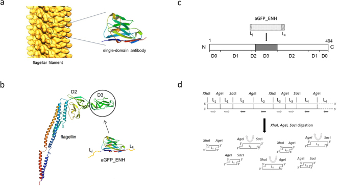
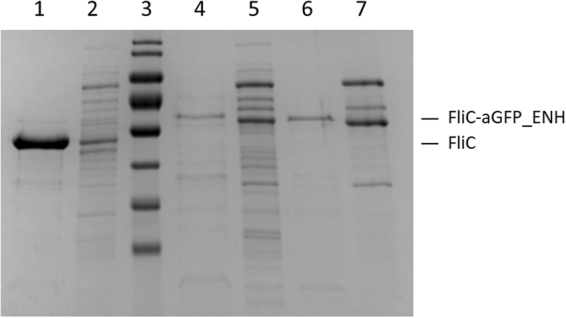
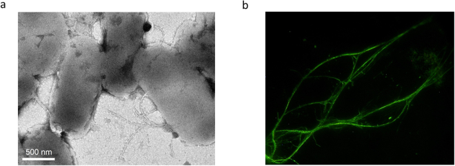
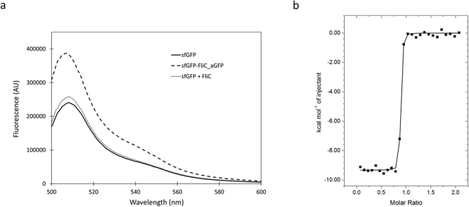
In this work we addressed the problem how to fabricate self-assembling tubular nanostructures displaying target recognition functionalities. Bacterial flagellar filaments, composed of thousands of flagellin subunits, were used as scaffolds to display single-domain antibodies (nanobodies) on their surface. As a representative example, an anti-GFP nanobody was successfully inserted into the middle part of flagellin replacing the hypervariable surface-exposed D3 domain. A novel procedure was developed to select appropriate linkers required for functional internal insertion. Linkers of various lengths and conformational properties were chosen from a linker database and they were randomly attached to both ends of an anti-GFP nanobody to facilitate insertion. Functional fusion constructs capable of forming filaments on the surface of flagellin-deficient host cells were selected by magnetic microparticles covered by target GFP molecules and appropriate linkers were identified. TEM studies revealed that short filaments of 2-900 nm were formed on the cell surface. ITC and fluorescent measurements demonstrated that the fusion protein exhibited high binding affinity towards GFP. Our approach allows the development of functionalized flagellar nanotubes against a variety of important target molecules offering potential applications in biosensorics and bio-nanotechnology.
Conflict of interest statement
The authors declare no competing interests.
Figures
Similar articles
-
A polymerizable GFP variant.Protein Eng Des Sel. 2012 Apr;25(4):153-7. doi: 10.1093/protein/gzs003. Epub 2012 Feb 2. Protein Eng Des Sel. 2012. PMID: 22301275
-
Construction of a xylanase A variant capable of polymerization.PLoS One. 2011;6(9):e25388. doi: 10.1371/journal.pone.0025388. Epub 2011 Sep 23. PLoS One. 2011. PMID: 21966517 Free PMC article.
-
Structural basis for stabilization of the hypervariable D3 domain of Salmonella flagellin upon filament formation.J Mol Biol. 2010 Nov 5;403(4):607-15. doi: 10.1016/j.jmb.2010.09.024. Epub 2010 Sep 22. J Mol Biol. 2010. PMID: 20868693
-
Nanobody-derived nanobiotechnology tool kits for diverse biomedical and biotechnology applications.Int J Nanomedicine. 2016 Jul 21;11:3287-303. doi: 10.2147/IJN.S107194. eCollection 2016. Int J Nanomedicine. 2016. PMID: 27499623 Free PMC article. Review.
-
The archaeabacterial flagellar filament: a bacterial propeller with a pilus-like structure.J Mol Microbiol Biotechnol. 2006;11(3-5):208-20. doi: 10.1159/000094055. J Mol Microbiol Biotechnol. 2006. PMID: 16983196 Review.
Cited by
-
Exploring cellular biochemistry with nanobodies.J Biol Chem. 2020 Nov 6;295(45):15307-15327. doi: 10.1074/jbc.REV120.012960. Epub 2020 Aug 31. J Biol Chem. 2020. PMID: 32868455 Free PMC article. Review.
-
Photobodies: Light-Activatable Single-Domain Antibody Fragments.Angew Chem Int Ed Engl. 2020 Jan 20;59(4):1506-1510. doi: 10.1002/anie.201912286. Epub 2019 Dec 12. Angew Chem Int Ed Engl. 2020. PMID: 31755215 Free PMC article.
-
Grating-coupled interferometry reveals binding kinetics and affinities of Ni ions to genetically engineered protein layers.Sci Rep. 2020 Dec 17;10(1):22253. doi: 10.1038/s41598-020-79226-w. Sci Rep. 2020. PMID: 33335217 Free PMC article.
-
Flagellin-based electrochemical sensing layer for arsenic detection in water.Sci Rep. 2021 Feb 10;11(1):3497. doi: 10.1038/s41598-021-83053-y. Sci Rep. 2021. PMID: 33568718 Free PMC article.
-
Extreme thermal stability of the antiGFP nanobody - GFP complex.BMC Res Notes. 2023 Jun 20;16(1):110. doi: 10.1186/s13104-023-06382-3. BMC Res Notes. 2023. PMID: 37340471 Free PMC article.
References
-
- Vonderviszt, F. & Namba, K. Structure, function and assembly of flagellar axial proteins in Fibrous Proteins (ed. Scheibel, T.) 58–76 (Landes Biosciences, 2008).
-
- Kumara MT, Tripp BC, Muralidharan S. Self-assembly of metal nanoparticles and nanotubes on bioengineered flagella scaffolds. Chem. Mater. 2007;19:2056–2064. doi: 10.1021/cm062178b. - DOI
Publication types
MeSH terms
Substances
LinkOut - more resources
Full Text Sources
Other Literature Sources
Research Materials
