

The role of cytokines and chemokines in the microenvironment of the blood-brain barrier in leukemia central nervous system metastasis
- PMID: 29483784
- PMCID: PMC5815469
- DOI: 10.2147/CMAR.S152419
The role of cytokines and chemokines in the microenvironment of the blood-brain barrier in leukemia central nervous system metastasis
Abstract
Aim: Central nervous system (CNS) metastasis is a major obstacle in the treatment of leukemia, and the underlying mechanisms of leukemia CNS metastasis are not fully understood. The present study is an investigation of the role of the CNS microenvironment in leukemia CNS metastasis.
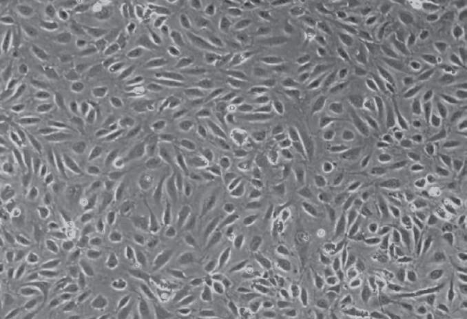
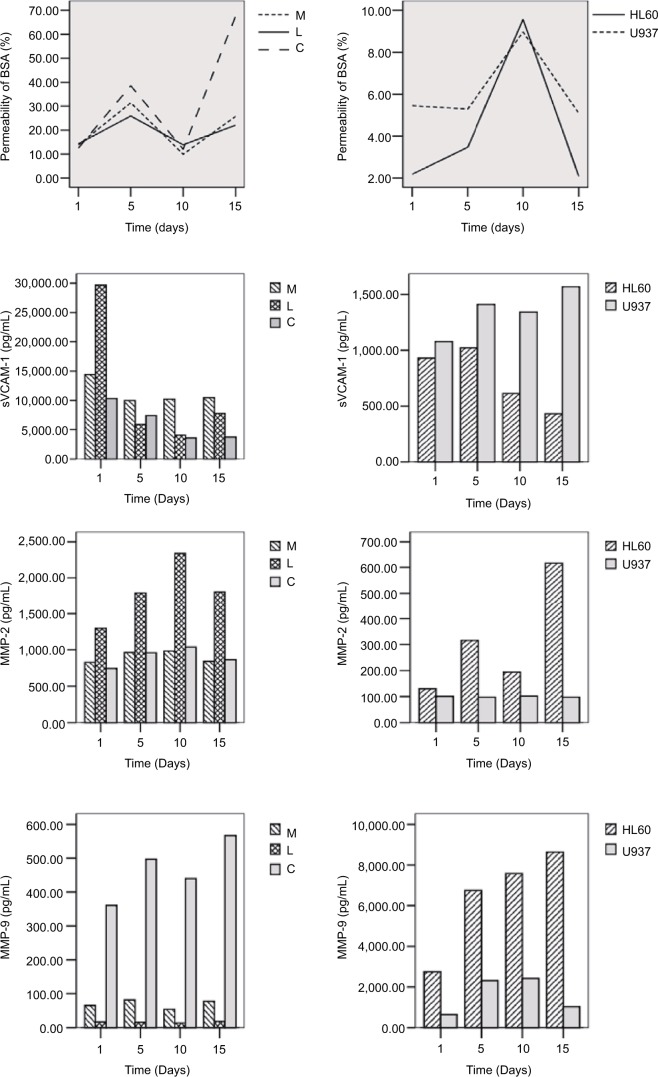
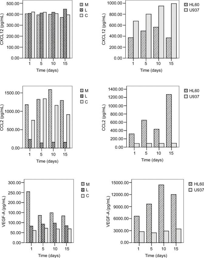
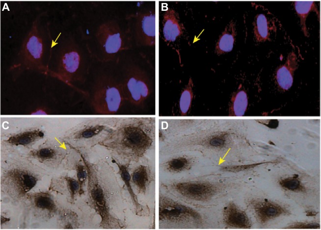
Methods: Analog blood-brain barrier (BBB) was set by coculturing human brain microvascular endothelial cells (HBMVECs) and leukemia cells (U937 and IL-60), as well as HBMVECs and sera from leukemia patients, in vitro. The permeability of the HBMVEC monolayer and the levels of tight junction proteins, cytokines and chemokines (C&Ckines) were measured.
Results: The permeability of HBMVECs increased when cocultured with leukemia sera. The expression of C&Ckines was significantly upregulated in HBMVECs cocultured with leukemia sera or leukemia cells, compared to the normal sera (P<0.05, respectively). Specifically, significantly higher levels of vascular endothelial growth factor A (VEGF-A) and matrix metalloprotease 9 (MMP-9) were found in HBMVECs and leukemia cells/sera coculturing systems.
Conclusion: Both leukemia cells and the molecules in leukemia sera play an important role in leukemia CNS metastasis. VEGF-A and MMPs may be the main factors resulting in the degradation of the BBB and inducing the CNS migration of leukemia cells.
Keywords: CNS leukemia; IL-60; U937; chemokine; cytokine.
Conflict of interest statement
Disclosure The authors report no conflicts of interest in this work.
Figures
Similar articles
-
Does VEGF secreted by leukemic cells increase the permeability of blood-brain barrier by disrupting tight-junction proteins in central nervous system leukemia?Med Hypotheses. 2011 May;76(5):618-21. doi: 10.1016/j.mehy.2010.12.001. Epub 2011 Mar 12. Med Hypotheses. 2011. PMID: 21398042
-
Matrix metalloproteinase-2 and -9 secreted by leukemic cells increase the permeability of blood-brain barrier by disrupting tight junction proteins.PLoS One. 2011;6(8):e20599. doi: 10.1371/journal.pone.0020599. Epub 2011 Aug 17. PLoS One. 2011. PMID: 21857898 Free PMC article.
-
[Critical roles of matrix metalloproteinases secreted by leukemic cells in the pathogenesis of central nervous system leukemia].Zhonghua Xue Ye Xue Za Zhi. 2016 Dec 14;37(12):1070-1076. doi: 10.3760/cma.j.issn.0253-2727.2016.12.012. Zhonghua Xue Ye Xue Za Zhi. 2016. PMID: 28088972 Free PMC article. Chinese.
-
Blood-brain barrier disruption in multiple sclerosis.Mult Scler. 2003 Dec;9(6):540-9. doi: 10.1191/1352458503ms965oa. Mult Scler. 2003. PMID: 14664465 Review.
-
The blood-brain and the blood-cerebrospinal fluid barriers: function and dysfunction.Semin Immunopathol. 2009 Nov;31(4):497-511. doi: 10.1007/s00281-009-0177-0. Epub 2009 Sep 25. Semin Immunopathol. 2009. PMID: 19779720 Review.
Cited by
-
The Impact of Small Extracellular Vesicles on Lymphoblast Trafficking across the Blood-Cerebrospinal Fluid Barrier In Vitro.Int J Mol Sci. 2020 Jul 31;21(15):5491. doi: 10.3390/ijms21155491. Int J Mol Sci. 2020. PMID: 32752027 Free PMC article.
-
Loss of IRF7 accelerates acute myeloid leukemia progression and induces VCAM1-VLA-4 mediated intracerebral invasion.Oncogene. 2022 Apr;41(16):2303-2314. doi: 10.1038/s41388-022-02233-w. Epub 2022 Mar 7. Oncogene. 2022. PMID: 35256780 Free PMC article.
-
Effects of VEGFR1+ hematopoietic progenitor cells on pre-metastatic niche formation and in vivo metastasis of breast cancer cells.J Cancer Res Clin Oncol. 2019 Feb;145(2):411-427. doi: 10.1007/s00432-018-2802-6. Epub 2018 Nov 27. J Cancer Res Clin Oncol. 2019. PMID: 30483898 Free PMC article.
-
Cerebrospinal fluid interleukin-6 is a potential diagnostic biomarker for central nervous system involvement in adult acute myeloid leukemia.Front Oncol. 2022 Dec 1;12:1013781. doi: 10.3389/fonc.2022.1013781. eCollection 2022. Front Oncol. 2022. PMID: 36531024 Free PMC article.
-
Central Nervous System Involvement in Adults with Acute Leukemia: Diagnosis, Prevention, and Management.Curr Oncol Rep. 2022 Apr;24(4):427-436. doi: 10.1007/s11912-022-01220-4. Epub 2022 Feb 10. Curr Oncol Rep. 2022. PMID: 35141858 Review.
References
-
- Si MY, Fan ZC, Li YZ, Chang XL, Xie QD, Jiao XY. The prognostic significance of serum and cerebrospinal fluid MMP-9, CCL2 and sVCAM-1 in leukemia CNS metastasis. J Neurooncol. 2015;122(2):229–244. - PubMed
-
- Tabouret E, Bauchet L, Carpentier AF. Brain metastases epidemiology and biology. Bull Cancer. 2013;100(1):57–62. - PubMed
-
- Gomes HR. Cerebrospinal fluid approach on neuro-oncology. Arq Neuropsiquiatr. 2013;71(9B):677–680. - PubMed
-
- Ransohoff RM, Kivisakk P, Kidd G. Three or more routes for leukocyte migration into the central nervous system. Nat Rev Immunol. 2003;3(7):569–581. - PubMed
-
- Abbott NJ, Patabendige AA, Dolman DE, et al. Structure and function of the blood-brain barrier. Neurobiol Dis. 2010;37(1):13–25. - PubMed
LinkOut - more resources
Full Text Sources
Other Literature Sources
Miscellaneous
