. 2018 Jan 2:2018:2870503.
doi: 10.1155/2018/2870503. eCollection 2018.

Preventive Effects of Velvet Antler (Cervus elaphus) against Lipopolysaccharide-Induced Acute Lung Injury in Mice by Inhibiting MAPK/NF- κ B Activation and Inducing AMPK/Nrf2 Pathways

Affiliations

Preventive Effects of Velvet Antler (Cervus elaphus) against Lipopolysaccharide-Induced Acute Lung Injury in Mice by Inhibiting MAPK/NF- κ B Activation and Inducing AMPK/Nrf2 Pathways

Jui-Shu Chang et al. Evid Based Complement Alternat Med. .

Abstract

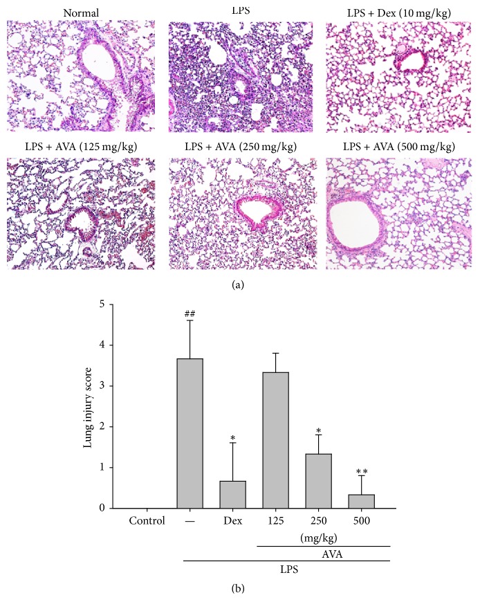
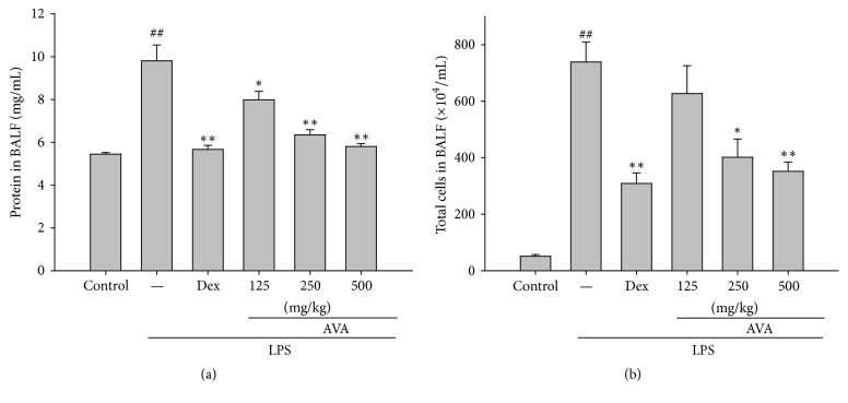
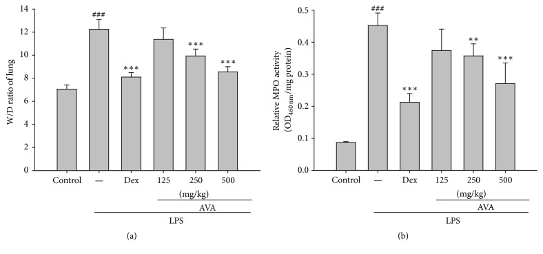
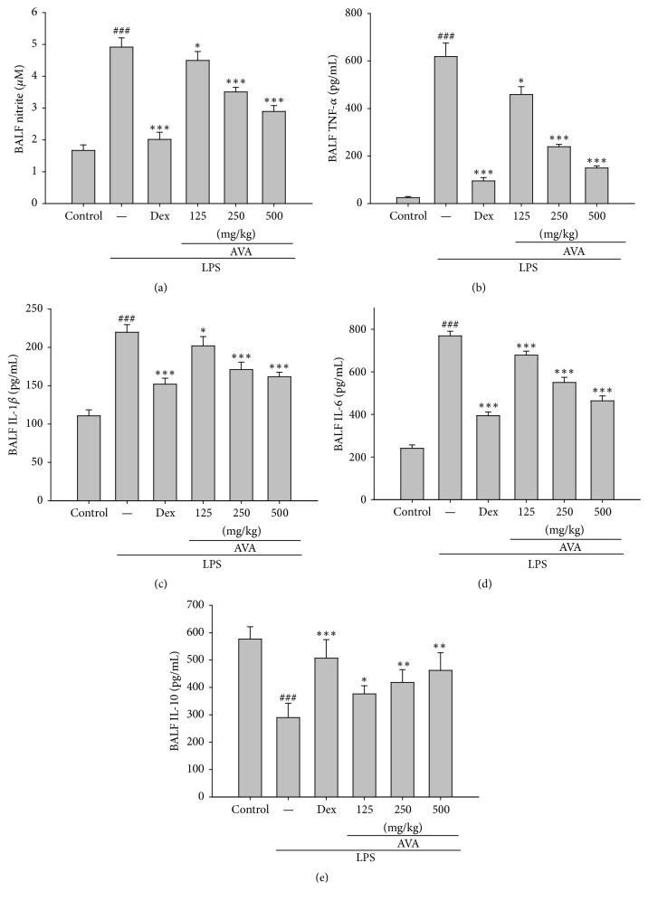
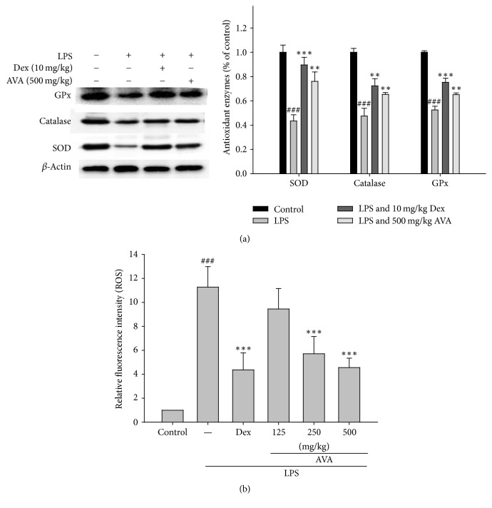
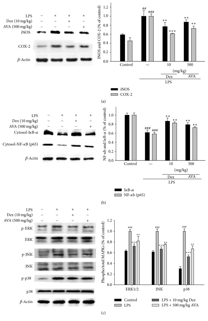
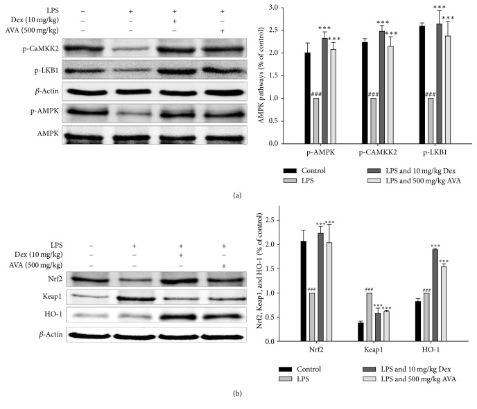
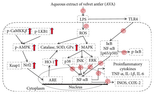
Velvet antler (Cervus elaphus) is a typical traditional animal medicine. It is considered to have various pharmacological effects including stimulation of the immune system, increase in the physical strength, and enhancement of sexual function. This paper aims to investigate the aqueous extract of velvet antler (AVA) in the mouse models of LPS-induced ALI. Inhibition of NO, TNF-α, IL-1β, IL-6, and IL-10 productions contributes to the attenuation of LPS-induced lung inflammation by AVA. A 5-day pretreatment of AVA prevented histological alterations and enhanced antioxidant enzyme activity in lung tissues. AVA significantly reduced the material (total number of cells and proteins) in the BALF. Western blot analysis revealed that the expression of iNOS and COX-2 and phosphorylation of IκB-α and MAPKs proteins are blocked in LPS-stimulated macrophages as well as LPS-induced lung injury in mice. Consistent with this concept, the phosphorylation of CaMKKβ, LKB1, AMPK, Nrf2, and HO-1 was activated after AVA treatment. The results from this study indicate AVA has anti-inflammatory effects in vivo and AVA is a potential model for the development of health food. In addition, its pathways may be at least partially associated with inhibiting MAPK/NF-κB activation and upregulating AMPK/Nrf2 pathways and the regulation of antioxidant enzyme activity.


Figures

Figure 1
Figure 1
The effects of the aqueous extract of velvet antler (AVA) on histopathological changes in the lung (a) and on the severity of lung injury were analyzed using the lung injury scoring system (b) in LPS-induced ALI mice. Mice were sacrificed 6 h after LPS stimulation. The right lungs were excised and embedded in 10% formalin, sectioned, and stained with H&E (magnification ×400). Images are representative of three experiments. Dex: dexamethasone. The data are presented as the mean ± SD for three different experiments performed in triplicate. ##Compared with the control group. p < 0.05 and ∗∗ p < 0.01 compared with the LPS-alone group.
Figure 2
Figure 2
Effects of AVA on the total protein level (a) and total cell number (b) in the BALF of LPS-induced ALI mice. Mice were given AVA (125, 250, and 500 mg/kg) by intraperitoneal injection 1 h before challenge with LPS. The BALF was collected 6 h after LPS challenge. Cell counts were assessed using a hemocytometer. Each value represents the mean ± SD. ## p < 0.01 compared with the control group. p < 0.05 and ∗∗ p < 0.01 compared with the LPS group (one-way ANOVA followed by Scheffe's multiple range test).
Figure 3
Figure 3
The effects of AVA on the lung W/D ratio (a) and myeloperoxidase activity (b) in LPS-induced ALI mice. Mice were given sclareol 1 h prior to intratracheal (i.t.) administration of LPS. The lung W/D ratio (a) and myeloperoxidase activity (b) were determined 6 h after LPS challenge. Each value represents the mean ± SD. ### p < 0.001 compared with the control group. ∗∗ p < 0.01 and ∗∗∗ p < 0.001 compared with the LPS group (one-way ANOVA followed by Scheffe's multiple range test).
Figure 4
Figure 4
The effect of AVA on NO (a), TNF-α (b), IL-1β (c), IL-6 (d), and IL-10 (f) expression in mice with ALI. Mice were given AVA (125, 250, and 500 mg/kg) via intraperitoneal injection 1 h before challenge with LPS. The BALF was collected 6 h following LPS challenge to analyze the inflammatory cytokines NO, TNF-α, IL-1β, IL-6, and IL-10. Each value represents the mean ± SD. ### p < 0.001 compared with the control group. p < 0.05, ∗∗ p < 0.01, and ∗∗∗ p < 0.001 compared with the LPS group (one-way ANOVA followed by Scheffe's multiple range test).
Figure 5
Figure 5
The antioxidant enzyme activity (a) and ROS production (b) of AVA in lungs from mice with ALI. Tissue homogenates were prepared and subjected to western blotting using antibodies specific for SOD, catalase, and GPx. β-Actin was used as an internal control. The BALF was collected 6 h following LPS challenge to analyze ROS production. The data are presented as the mean ± SD for three different experiments performed in triplicate. ###Compared with the control group. ∗∗ p < 0.01 and ∗∗∗ p < 0.001 compared with the LPS-alone group.
Figure 6
Figure 6
The inhibition of iNOS, COX-2 (a), IκB-α, NF-κB (b), and MAPK protein (c) expression by AVA in LPS-induced ALI mice. Tissue homogenates were prepared and subjected to western blotting using antibodies specific for iNOS, COX-2, IκB-α, NF-κB, and MAPK. The values under each lane indicate the relative band intensities normalized to β-actin. The data are presented as the mean ± SD for three different experiments performed in triplicate. ###Compared with the control group. ∗∗ p < 0.01 and ∗∗∗ p < 0.001 compared with the LPS-alone group.
Figure 7
Figure 7
The inhibition of CaMKKβ, LKB1, and AMPK (a) and HO-1, Nrf2, and Keap1 (b) expression by AVA in LPS-induced ALI mice. Tissue homogenates were prepared and subjected to western blotting using antibodies specific for p-CaMKKβ, p-LKB1, p-AMPK, AMPK, HO-1, Nrf2, and Keap1. The values under each lane indicate the relative band intensities normalized to β-actin. The data are presented as the mean ± SD for three different experiments performed in triplicate. ###Compared with the control group. ∗∗∗ p < 0.001 compared with the LPS-alone group.
Figure 8
Figure 8
The schemes of the mechanism for the protective effect of AVA on LPS-induced inflammation.

References

    1. Li K.-C., Ho Y.-L., Hsieh W.-T., Huang S.-S., Chang Y.-S., Huang G.-J. Apigenin-7-glycoside prevents LPS-induced acute lung injury via downregulation of oxidative enzyme expression and protein activation through inhibition of MAPK phosphorylation. International Journal of Molecular Sciences. 2015;16(1):1736–1754. doi: 10.3390/ijms16011736. - DOI - PMC - PubMed
    1. Huang G.-J., Deng J.-S., Chen C.-C., et al. Methanol extract of Antrodia camphorata protects against lipopolysaccharide-induced acute lung injury by suppressing NF-κB and MAPK pathways in mice. Journal of Agricultural and Food Chemistry. 2014;62(23):5321–5329. doi: 10.1021/jf405113g. - DOI - PubMed
    1. Matthay M. A., Zimmerman G. A. Acute lung injury and the acute respiratory distress syndrome: Four decades of inquiry into pathogenesis and rational management. American Journal of Respiratory Cell and Molecular Biology. 2005;33(4):319–327. doi: 10.1165/rcmb.F305. - DOI - PMC - PubMed
    1. Tsai C. L., Lin Y. C., Wang H. M., et al. Baicalein, an active component of Scutellaria baicalensis, protects against lipopolysaccharide-induced acute lung injury in rats. Journal of Ethnopharmacology. 2014;153(1):197–206. - PubMed
    1. Yao J., Pan D., Zhao Y., et al. Wogonin prevents lipopolysaccharide-induced acute lung injury and inflammation in mice via peroxisome proliferator-activated receptor γ-mediated attenuation of the nuclear factor-κβ pathway. The Journal of Immunology. 2014;143(2):241–257. doi: 10.1111/imm.12305. - DOI - PMC - PubMed