

Overexpression of MICAL2, a novel tumor-promoting factor, accelerates tumor progression through regulating cell proliferation and EMT
- PMID: 29483957
- PMCID: PMC5820919
- DOI: 10.7150/jca.22355
Overexpression of MICAL2, a novel tumor-promoting factor, accelerates tumor progression through regulating cell proliferation and EMT
Abstract
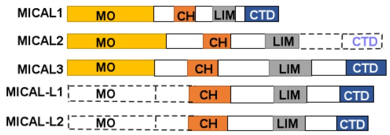
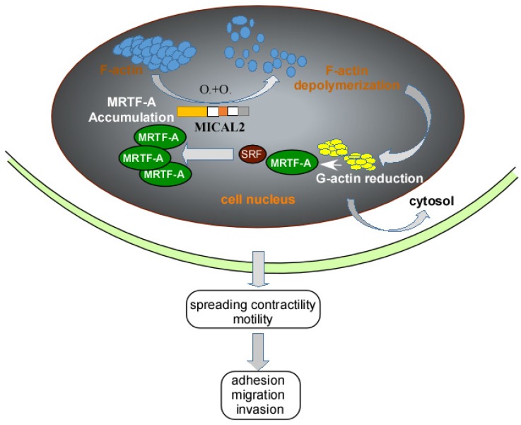
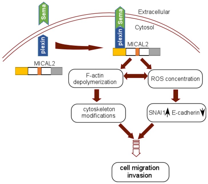
Molecule interacting with CasL 2 (MICAL2), a microtubule associated monooxygenase, is involved in cell growth, axon guidance, vesicle trafficking and apoptosis. Recent studies have demonstrated that MICAL2 is highly expressed in tumor and accelerates tumor progression and it is deemed to be a novel tumor-promoting factor. MICAL2 overexpression increases cell proliferation to accelerate tumor growth, and MICAL2 also promotes epithelial-mesenchymal transition (EMT)-related proteins to increase cancer cell metastasis. On mechanism, MICAL2 induces EMT by regulating SRF (serum response factor)/MRTF-A (myocardin related transcription factor A) signaling, Semaphorin/Plexin pathway and inducing ROS (Reactive oxygen species) production. In the present review, we introduced MICAL family, expatiated the structure and functions of MICALs, and summarized the mechanisms of MICAL2 involving tumor progression. The challenges and perspectives for MICAL2 in tumor are also discussed.
Keywords: MICAL2; cancer; epithelial-mesenchymal transition; metastasis..
Conflict of interest statement
Competing Interests: The authors have declared that no competing interest exists.
Figures
Similar articles
-
The role of MICAL2 in cancer progression: mechanisms, challenges, and therapeutic potential.Hum Cell. 2025 Apr 16;38(3):89. doi: 10.1007/s13577-025-01212-z. Hum Cell. 2025. PMID: 40240704 Review.
-
MICAL2 Is a Super Enhancer Associated Gene that Promotes Pancreatic Cancer Growth and Metastasis.bioRxiv [Preprint]. 2024 Jun 30:2024.06.26.600548. doi: 10.1101/2024.06.26.600548. bioRxiv. 2024. PMID: 38979336 Free PMC article. Preprint.
-
MICAL2 is a novel human cancer gene controlling mesenchymal to epithelial transition involved in cancer growth and invasion.Oncotarget. 2016 Jan 12;7(2):1808-25. doi: 10.18632/oncotarget.6577. Oncotarget. 2016. PMID: 26689989 Free PMC article.
-
MICAL2 Promotes Proliferation and Migration of Glioblastoma Cells Through TGF-β/p-Smad2/EMT-Like Signaling Pathway.Front Oncol. 2021 Nov 12;11:735180. doi: 10.3389/fonc.2021.735180. eCollection 2021. Front Oncol. 2021. PMID: 34868922 Free PMC article.
-
MICAL flavoprotein monooxygenases: structure, function and role in semaphorin signaling.Adv Exp Med Biol. 2007;600:38-51. doi: 10.1007/978-0-387-70956-7_4. Adv Exp Med Biol. 2007. PMID: 17607945 Review.
Cited by
-
Loci associated with conception rate in crossbred beef heifers.PLoS One. 2020 Apr 9;15(4):e0230422. doi: 10.1371/journal.pone.0230422. eCollection 2020. PLoS One. 2020. PMID: 32271764 Free PMC article.
-
The Role of Epicardial Adipose Tissue-Derived MicroRNAs in the Regulation of Cardiovascular Disease: A Narrative Review.Biology (Basel). 2023 Mar 25;12(4):498. doi: 10.3390/biology12040498. Biology (Basel). 2023. PMID: 37106699 Free PMC article. Review.
-
High MICAL1 expression correlates with cancer progression and immune infiltration in renal clear cell carcinoma.BMC Cancer. 2022 Dec 27;22(1):1355. doi: 10.1186/s12885-022-10462-1. BMC Cancer. 2022. PMID: 36575439 Free PMC article.
-
MICAL-L2 Is Essential for c-Myc Deubiquitination and Stability in Non-small Cell Lung Cancer Cells.Front Cell Dev Biol. 2021 Jan 14;8:575903. doi: 10.3389/fcell.2020.575903. eCollection 2020. Front Cell Dev Biol. 2021. PMID: 33520979 Free PMC article.
-
LncRNA MYO16-AS1 and MICAL2 axis sustains proliferation and migration in ovarian cancer cells and unveils a therapeutic vulnerability in patient-derived tumor organoids.Noncoding RNA Res. 2025 Jul 23;15:74-84. doi: 10.1016/j.ncrna.2025.07.005. eCollection 2025 Dec. Noncoding RNA Res. 2025. PMID: 40822859 Free PMC article.
References
-
- Suzuki T, Nakamoto T, Ogawa S, Seo S, Matsumura T, Tachibana K. et al. MICAL, a novel CasL interacting molecule, associates with vimentin. J Biol Chem. 2002;277:14933–41. - PubMed
-
- Xue Y, Kuok C, Xiao A, Zhu Z, Lin S, Zhang B. Identification and expression analysis of mical family genes in zebrafish. J Genet Genomics. 2010;37:685–93. - PubMed
-
- Ashida S, Furihata M, Katagiri T, Tamura K, Anazawa Y, Yoshioka H. et al. Expression of novel molecules, MICAL2-PV (MICAL2 prostate cancer variants), increases with high Gleason score and prostate cancer progression. Clin Cancer Res. 2006;12:2767–73. - PubMed
-
- Giordano S, Corso S, Conrotto P, Artigiani S, Gilestro G, Barberis D. et al. The semaphorin 4D receptor controls invasive growth by coupling with Met. Nat Cell Biol. 2002;4:720–4. - PubMed
Publication types
LinkOut - more resources
Full Text Sources
Other Literature Sources
Miscellaneous
