

Advanced Pretibial Melanoma (APM): Clinicians Behaviour As Triggering Factor?
- PMID: 29483985
- PMCID: PMC5816319
- DOI: 10.3889/oamjms.2018.003
Advanced Pretibial Melanoma (APM): Clinicians Behaviour As Triggering Factor?
Abstract
Background: Pigmented lesions represent a broad spectrum of clinical conditions, both benign and malignant. The precise diagnosis is often a challenge, while the clinical diagnostic criteria could be misleading, as a result of the frequently atypical presentation of otherwise completely benign in nature lesions. The variety of therapeutic options for benign pigmented lesions including shave curettage, local laser destruction, electrocoagulation removal could sound enticingly both for the physician and patient, but they destroy the possibility for histological examination and provide a deceptively feeling of calm, that the problem is solved. If there is even a minimum chance for misdiagnosis, the risk could be a human life. Furthermore, a simple surgical excision could provide total resolution of the problem, with correct histological verification and further therapeutic measurements, if needed.
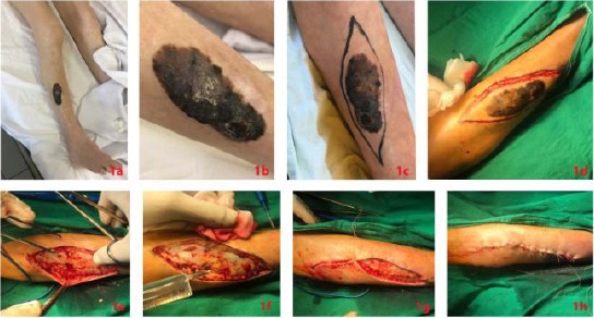
Case report: We present a case of a patient, with advanced pretibial melanoma with multiple lung metastases, misdiagnosed as a seborrheic keratosis, treated with shave-curettage 6 months earlier, as we want to emphasize the importance of the correct therapeutic method in all cases with pigmented lesions with unknown origin, in order to minimize the risk of dramatic consequences of misdiagnosis of melanoma. So, we want to ask you- is this risk justified?
Conclusion: So, we want to ask you - is this risk justified?
Keywords: imitation; melanoma; outcome; risk behaviour; seborrheic keratosis; surgery.
Figures
References
-
- Blum A, Kreusch J, Stolz W, Haenssle H, Braun R, Hofmann-Wellenhof R, Tschandl P, Zalaudek I, Kittler H. Dermoscopy for malignant and benign skin tumours:Indication and standardized terminology. Hautarzt. 2017;68(8):653–73. https://doi.org/10.1007/s00105-017-4013-5 PMid:28721529. - PubMed
-
- Carli P, De Giorgi V, Crocetti E, Mannone F, Massi D, Chiarugi A, Giannotti B. Improvement of malignant/benign ratio in excised melanocytic lesions in the ‘dermoscopy era’:a retrospective study 1997-2001. Br J Dermatol. 2004;150(4):687–92. https://doi.org/10.1111/j.0007-0963.2004.05860.x PMid:15099364. - PubMed
-
- Marino ML, Carrera C, Marchetti MA, Marghoob AA. Practice Gaps in Dermatology:Melanocytic Lesions and Melanoma. Dermatol Clin. 2016;34(3):353–62. https://doi.org/10.1016/j.det.2016.03.003 PMid:27363893. - PubMed
-
- de Giorgi V, Savarese I, Rossari S, Gori A, Grazzini M, Crocetti E, Sara Longo A, Oranges T, Massi D. Features of small melanocytic lesions:does small mean benign? A clinical-dermoscopic study. Melanoma Res. 2012;22(3):252–6. https://doi.org/10.1097/CMR.0b013e3283527430 PMid:22430838. - PubMed
-
- Schein O, Westreich M, Shalom A. Effect of dermoscopy on diagnostic accuracy of pigmented skin lesions emphasizing malignant melanoma. Harefuah. 2009;148(12):820–3, 855. PMid:20088434. - PubMed
Publication types
LinkOut - more resources
Full Text Sources
Other Literature Sources
Research Materials