Case Reports
. 2017 Jul 31;12(4):672-677.
doi: 10.1016/j.radcr.2017.06.005. eCollection 2017 Dec.

Bifid sternum in a young woman: Multimodality imaging features

Affiliations
Case Reports

Bifid sternum in a young woman: Multimodality imaging features

Guglielmo Manenti et al. Radiol Case Rep. .

Abstract

Bifid sternum is a rare fusion anomaly of the chest wall that accounts for 0.15% of all chest deformities and may be associated with cardiac or vascular anomalies. It is usually diagnosed and surgically corrected at birth or within the first month of life. Being a diagnosis made during the neonatal period, computed tomography scan and magnetic resonance imaging are not often performed; not so many cases in literature have been studied with II level diagnostic imaging, such as computed tomography or magnetic resonance. We describe a case of bifid sternum, rarely diagnosed in adults, discovered in a 21-year-old woman who came to our Diagnostic Imaging Department to perform a chest magnetic resonance after a chest X-ray.

Keywords: Bifid; CT; Cleft; MRI; Sternum.


Figures

Fig. 1
Fig. 1
Twenty-one-year-old woman with bifid sternum. Findings: Double-projection chest X-ray. (A) Figure demonstrates no pathologic findings in posteroanterior (PA) projection. (B) Figure demonstrates an abnormal radiolucent area (red arrow) in the upper part of the thoracic wall in latero-lateral (L-L) projection. The patient shows a bony defect in the central upper part of her chest wall, more noticeable during inspiration, without alterations in the laboratory tests, echocardiography, and electrocardiogram. Technique: PA and L-L projections of chest X-ray.
Fig. 2
Fig. 2
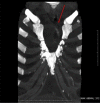
Twenty-one-year-old woman with bifid sternum. Findings: Noncontrast chest coronal magnetic resonance imaging (MRI). (A and B) Figures demonstrate the sternal fusion defect with an incomplete superior cleft of the manubrium and the body of the sternum (red arrows). This is a rare fusion anomaly of the chest wall that occurs in 0.15% of all chest deformities. It is usually diagnosed and corrected surgically at birth or within the first month of life, so it is rarely diagnosed in adulthood. Technique: 1.5 Tesla General Electric (GE) software version 15 (GE Healthcare, Milwaukee, WI); noncontrast chest coronal T1-weighted (W) turbo spin echo (TSE) (TR 613 ms, TE 8 ms, slice thickness 3 mm, skip 1 mm).
Fig. 3
Fig. 3
Twenty-one-year-old woman with bifid sternum. Findings: Noncontrast chest axial MRI demonstrates lack of fusion of the sternal margins (red arrow), with the right ventricle directly in contact with the chest wall (yellow arrow). The bony defect in the central upper part of the chest was evident since birth and more noticeable during inspiration, but heart pulsations were not clinically evident on the chest wall. There were no alterations in the laboratory tests, echocardiography, and electrocardiogram. Technique: 1.5 Tesla General Electric (GE) software version 15 (GE Healthcare, Milwaukee, WI); noncontrast chest axial T1-weighted (W) turbo spin echo (TSE) (TR 613 ms, TE 8 ms, slice thickness 3 mm, skip 1 mm).
Fig. 4
Fig. 4
Twenty-one-year-old woman with bifid sternum. Findings: Noncontrast chest T2-weighted magnetic resonance imaging (MRI) on coronal plane shows bifid sternum with the incomplete superior cleft of the manubrium and the body of the sternum (red arrow). Technique: 1.5 Tesla General Electric (GE) software version 15 (GE Healthcare, Milwaukee, WI); noncontrast chest coronal T2-weighted (W) turbo spin echo (TSE) (TR 3500 ms, TE 100 ms, slice thickness 3 mm, skip 1 mm).
Fig. 5
Fig. 5
Twenty-one-year-old woman with bifid sternum. Findings: volume rendering reconstruction of noncontrast chest computed tomography (CT). (A and B) Figures show 2 views of cleft of the upper part of the sternum with “U-shape” variant. The superior sternal cleft may be “V-shaped” when the cleft reaches the xiphoid process, or “U-shaped,” with a bony bridge joining the 2 edges ending at the third or fourth costal cartilages, as seen in this case. Technique: CT GE (General Electric Company) LIGHTSPEED volumetric computed tomography 64 slices; noncontrast CT with volume rendering reconstruction (helical scan; 0.7-s rotation time; pitch 0.9; 120 kV; 200 mA; image thickness of 1.25 mm; bone plus filter).
Fig. 6
Fig. 6
Twenty-one-year-old woman with bifid sternum. Findings: maximum intensity projection-3-dimensional reconstruction of noncontrast chest computed tomography (CT) demonstrates the cleft of the upper part of the sternum (red arrow). This is a rare fusion anomaly of the chest wall that occurs in the 0.15% of all chest deformities. Technique: CT GE (General Electric Company) LIGHTSPEED volumetric computed tomography 64 slices; non-contrast CT with maximum intensity projection-3D reconstruction (helical scan; 0.7-s rotation time; pitch 0.9; 120 kV; 200 mA; image thickness of 1.25 mm; bone plus filter).

References

    1. Bayaroğulları H., Yengil E., Davran R., Ağlagül E., Karazincir S., Balcı A. Evaluation of the postnatal development of the sternum and sternal variations using multidetector CT. Diagn Interv Radiol. 2014;20(1):82–89. 24100061 PMID. - PMC - PubMed
    1. Torre M., Rapuzzi G., Carlucci M., Pio L., Jasonni V. Phenotypic spectrum and management of sternal cleft: literature review and presentation of a new series. Eur J Cardiothorac Surg. 2011;41(1):4–9. 21737294 PMID. - PMC - PubMed
    1. Das S.K., Jana P.K., Bairagya T.D., Ghoshal B. Bifid sternum. Lung India. 2012;29(1):73–75. 22345921 PMID. - PMC - PubMed
    1. Katranci A.O., Görk A.S., Rizalar R., Günaydin M., Aritürk E., Bernay F. Pentalogy of Cantrell. Indian J Pediatr. 1998;65(1):149–153. 10771959 PMID. - PubMed
    1. Ravitch M.M. WB Saunders; Philadelphia (PA): 1977. Congenital deformities of the chest wall and their operative correction. 9780721674797.

Publication types

LinkOut - more resources