. 2017 Nov 20;9(1):e1389360.
doi: 10.1080/19381980.2017.1389360. eCollection 2017.

Differential biological effects of dehydroepiandrosterone (DHEA) between mouse (B16F10) and human melanoma (BLM) cell lines

Affiliations

Differential biological effects of dehydroepiandrosterone (DHEA) between mouse (B16F10) and human melanoma (BLM) cell lines

Kumud Joshi et al. Dermatoendocrinol. .

Abstract

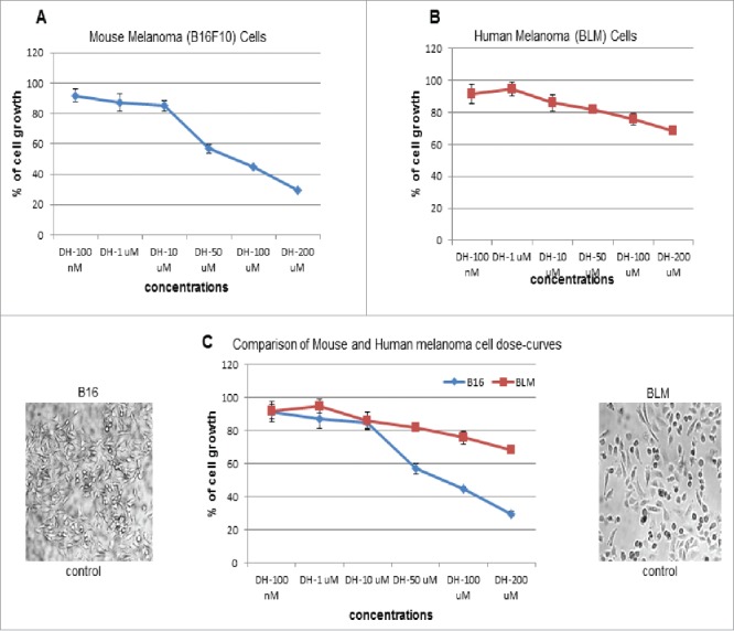
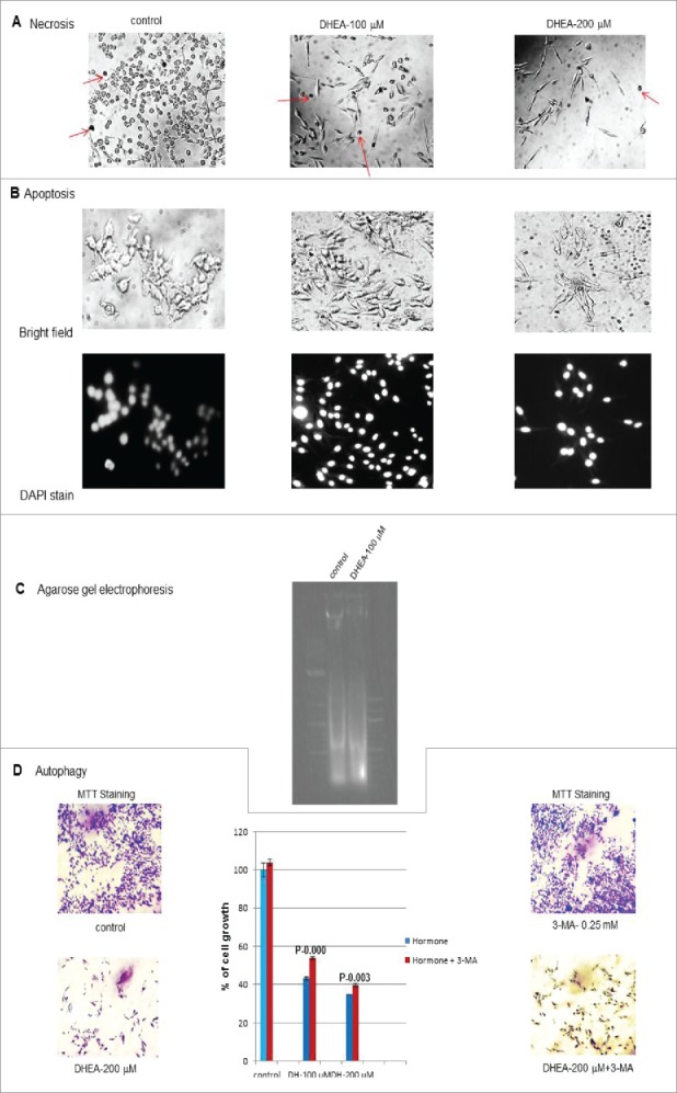
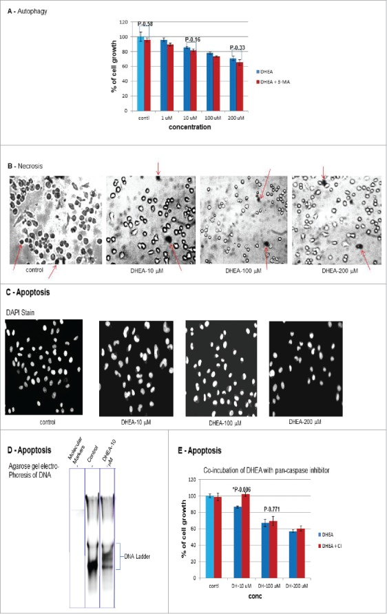
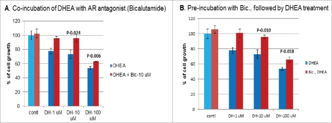
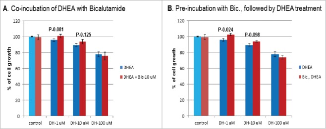
Dehydroepiandrosterone (DHEA) is a weak androgen and had been shown to have anti-cancer, anti-adipogenic and anti-inflammatory effects on mouse and other rodent models, but not on humans, suggesting a systemic level difference between mouse and human. Our previous study on DHEA biological functions involving a variety of cell lines, suggested that the functional differences between mouse and human existed even at the cellular level. Hence, using mouse and human melanoma cell models, in-vitro effects of DHEA on cell growth, mechanism of cell death and mechanism of DHEA action were studied. Results indicated a differential biological effects of DHEA between mouse and human melanoma cell lines. These in-vitro studies also suggested that the differential biological effects observed between these two cell lines could be due to the difference in the way DHEA was processed or metabolized inside the cell.

Keywords: Bicalutamide; DHEA; androgen receptor; apoptosis; autophagy; human melanoma (BLM) cell line; mouse melanoma (B16F10) cell line.


Figures

Figure 1.
Figure 1.
Comparison of Dose-response curves: Dose response studies were carried out with mouse and human melanoma cell lines starting from 100 nM up to 200 μM concentrations of DHEA. Cells were incubated with DHEA for 48 hrs. After 48 hrs of incubation, MTT assay was carried out to check cell growth. (A) Mouse melanoma (B16F10) cells showed a dose-dependent decrease in cell growth and significant inhibition (30%) at 200 μM concentration. (B) Human melanoma (BLM) cells showed a muffled response with mild inhibition of cell growth (69%) even at 200 μM concentration of DHEA. (C) When dose-response curves of both cell lines were compared, the difference in the response appeared after 10 μM concentration of DHEA.
Figure 2.
Figure 2.
Mechanism of mouse melanoma cell growth inhibition: DHEA treatment resulted in the inhibition of cell growth. The mechanism of inhibition of cell growth was investigated. (A) Necrosis: Necrosis as the cause of cell death was checked first, using 0.4% trypan blue dye. Dead cells would take up the dye and appear as purple colored cells under microscope. There was no difference in the number of stained (arrows point stained cells) cells between control and DHEA (100 μM, 200 μM) treated cells, suggesting necrosis was not the mechanism of cell death. (B) Apoptosis: Apoptosis or programed cell death as the mechanism was checked initially by staining the cells with DAPI (a fluorescent probe, which specifically stains nucleus) for change in nuclear shape due to condensation of chromatin. The nuclei were circular or oval shaped both in the control and DHEA treated cells, indicating no change in the shape of nucleus after DHEA treatment. (C) Agarose gel electrophoresis: DNA was harvested from untreated control and DHEA 100 μM treated cells. Fifteen μgm of DNA was loaded on 1.2% agarose gel to check for DNA ladder formation. There was no difference in the DNA pattern between control and DHEA 100 μM treated cells, suggesting apoptosis was not the mechanism of cell death. (D) Autophagy: Finally autophagy as the mechanism of cell death was checked by co-incubating cells with DHEA + 3-methyl adenine (0.25 mM). Three methyl adenine was able to partially rescue cell growth in DHEA 100 and 200 μM treated cells compared to plain DHEA treated cells.
Figure 3.
Figure 3.
Mechanism of inhibition of human melanoma (BLM) cell growth: (A) Autophagy: Since, mouse melanoma cell growth was inhibited by autophagy, it was decided to check autophagy first, as the cause of human melanoma cell death. Cells were co-incubated with DHEA or DHEA + 3-MA 2 mM (concentration of 3-MA was decided by trial and error (12)). After 48 hrs of incubation, cell growth was assessed by MTT assay. There was no difference in cell growth between plain DHEA treated or DHEA + 3-MA co-incubated cells, suggesting autophagy was not the mechanism of cell death. (B) Necrosis: Necrosis as the possible mechanism of inhibition of cell growth was checked by staining cells with 0.4% trypan blue. There was no difference in the number of stained cells (arrows point stained cells) between control and DHEA (10, 100 and 200 μM) treated cells, suggesting necrosis was not the mechanism of cell death. (C) Apoptosis: Apoptosis as the mechanism of cell death was checked first by staining cells with DAPI for change in nuclear shape. DAPI staining showed DHEA treated cells with sickle or crescent-moon shaped nuclei compared to circular or oval shaped nuclei of untreated control cells, suggesting apoptosis could be the mechanism of cell death. (D) DNA agarose gel electrophoresis: DNA was isolated from control and DHEA (10 μM) treated cells to check for DNA ladder formation in DHEA treated cells. Fifteen microgram of DNA was loaded on the gel and ran at 100 volts. The gel was stained with ethidium bromide and viewed under UV light. DHEA treated cells showed DNA ladder formation compared to untreated control cell DNA, suggesting apoptosis was the mechanism of cell death in human melanoma cells. (E) Incubation with pan-caspase inhibitor: In order to further confirm apoptosis, human melanoma cells were incubated with DHEA 10, 100 and 200 μM concentrations either alone or with 10 μM concentration of pan-caspase inhibitor (as higher concentration of caspase inhibitor inhibited cell growth by itself). As expected inhibition of caspase by caspase inhibitor (10 μM) along with DHEA 10 μM showed a complete recovery of cell growth. However, such complete or partial recovery of cell growth was not seen at 100 and 200 μM concentrations of DHEA. This could be explained because DHEA was at 10 and 20 times higher concentration when compared to pan caspase inhibitor concentration (10 μM) in these samples. So pan caspase inhibitor could not inhibit caspase activity at these high concentrations of DHEA.
Figure 4.
Figure 4.
Mechanism of DHEA action on mouse melanoma cells: As mechanism of cell death between mouse and human melanoma cells were different, it was decided to check whether the action of DHEA was mediated through androgen receptor. This assay was carried out in two ways. (A) First by co-incubating AR antagonist bicalutamide (10 μM) with DHEA at 1, 10, 100 μM concentrations for 48 hrs. There was an increase in cell growth at all the concentrations of DHEA + Bicalutamide co-incubated cells compared to DHEA alone incubated cells, indicating there was a competition between DHEA and bicalutamide for AR. (B) Secondly, mouse melanoma cells were pre-incubated with bicalutamide (10 μM) for 60 – 90 min and later DHEA was added. Mouse melanoma Cells showed an increase in cell growth at all the concentrations of DHEA treatment after bicalutamide pre-incubation compared to straight DHEA treatment, suggesting the action of DHEA was mediated through AR.
Figure 5.
Figure 5.
Mechanism of DHEA action on human melanoma cells: Same type of co-incubation and pre-incubation experiments were carried out with human melanoma cells. (A) Co-incubation of DHEA with bicalutamide (10 μM) did not show any increase in human melanoma cell growth compared to control cells, suggesting there was no competition between DHEA and bicalutamide for AR. (B) Similarly pre-incubation of cells with bicalutamide, followed by DHEA treatment did not increase the cell growth, again suggesting there was no competition between DHEA and bicalutamide for AR. So DHEA action was not mediated through AR.

Similar articles

Cited by

References

    1. Hall PF, Sozer CC, Eik-Nes KB. Formation of dehydroepiandrosterone during in vivo and in vitro biosynthesis of testosterone by testicular tissue. Endocrinol. 1964;74:35–43. doi:10.1210/endo-74-1-35. PMID:14114668. - DOI - PubMed
    1. Symington T, Duguid WP, Davidson JN. Effect of exogenous corticotropin on the histochemical pattern of the human adrenal cortex and a comparison with the changes during stress. JCEM, 1956;16:580–98. - PubMed
    1. Kalimi M, Regelson M (Eds) The biological role of dehydroepiandrosterone (DHEA). New York: Walter de gruyter; 1990.
    1. Baulieu EE. Dehydroepiandrosterone: A fountain of youth? JCEM. 1996;81(9):3147–51. - PubMed
    1. Khorram O. DHEA: a hormone with multiple effects. Current opinion in obstetrics and gynecology. 1996;8:351–4. doi:10.1097/00001703-199610000-00006. - DOI - PubMed

LinkOut - more resources