. 2018 Feb 12:5:6.
doi: 10.3389/fnut.2018.00006. eCollection 2018.

Improved Exercise Tolerance with Caffeine Is Associated with Modulation of both Peripheral and Central Neural Processes in Human Participants

Affiliations

Improved Exercise Tolerance with Caffeine Is Associated with Modulation of both Peripheral and Central Neural Processes in Human Participants

Joanna L Bowtell et al. Front Nutr. .

Abstract

Background: Caffeine has been shown to enhance exercise performance and capacity. The mechanisms remain unclear but are suggested to relate to adenosine receptor antagonism, resulting in increased central motor drive, reduced perception of effort, and altered peripheral processes such as enhanced calcium handling and extracellular potassium regulation. Our aims were to investigate how caffeine (i) affects knee extensor PCr kinetics and pH during repeated sets of single-leg knee extensor exercise to task failure and (ii) modulates the interplay between central and peripheral neural processes. We hypothesized that the caffeine-induced extension of exercise capacity during repeated sets of exercise would occur despite greater disturbance of the muscle milieu due to enhanced peripheral and corticospinal excitatory output, central motor drive, and muscle contractility.

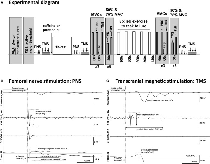
Methods: Nine healthy active young men performed five sets of intense single-leg knee extensor exercise to task failure on four separate occasions: for two visits (6 mg·kg-1 caffeine vs placebo), quadriceps 31P-magnetic resonance spectroscopy scans were performed to quantify phosphocreatine kinetics and pH, and for the remaining two visits (6 mg·kg-1 caffeine vs placebo), femoral nerve electrical and transcranial magnetic stimulation of the quadriceps cortical motor area were applied pre- and post exercise.

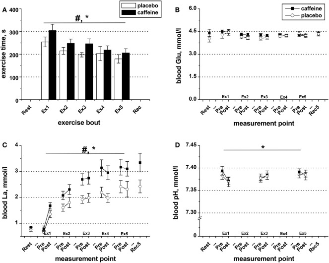
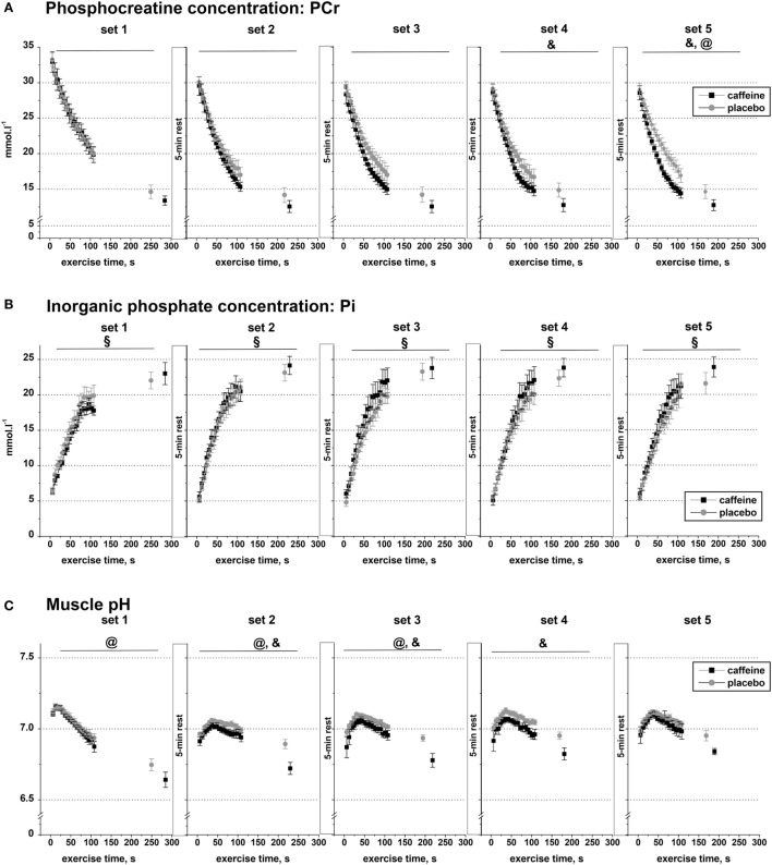
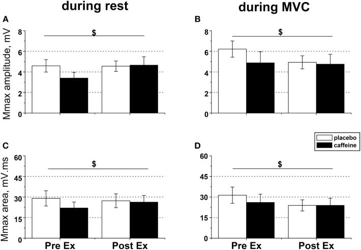
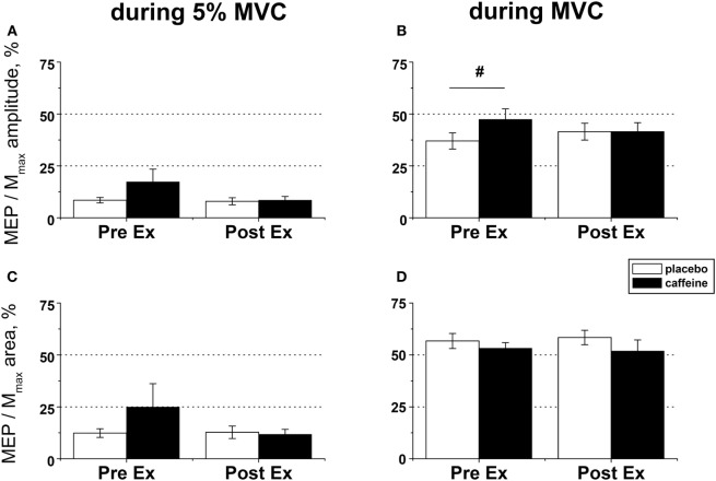
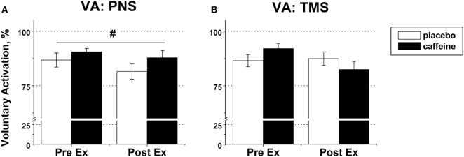
Results: The total exercise time was 17.9 ± 6.0% longer in the caffeine (1,225 ± 86 s) than in the placebo trial (1,049 ± 73 s, p = 0.016), and muscle phosphocreatine concentration and pH (p < 0.05) were significantly lower in the latter sets of exercise after caffeine ingestion. Voluntary activation (VA) (peripheral, p = 0.007; but not supraspinal, p = 0.074), motor-evoked potential (MEP) amplitude (p = 0.007), and contractility (contraction time, p = 0.009; and relaxation rate, p = 0.003) were significantly higher after caffeine consumption, but at task failure MEP amplitude and VA were not different from placebo. Caffeine prevented the reduction in M-wave amplitude that occurred at task failure (p = 0.039).

Conclusion: Caffeine supplementation improved high-intensity exercise tolerance despite greater-end exercise knee extensor phosphocreatine depletion and H+ accumulation. Caffeine-induced increases in central motor drive and corticospinal excitability were attenuated at task failure. This may have been induced by the afferent feedback of the greater disturbance of the muscle milieu, resulting in a stronger inhibitory input to the spinal and supraspinal motor neurons. However, causality needs to be established through further experiments.

Keywords: caffeine; central fatigue; fatigue; neuromuscular function; transcranial magnetic stimulation.


Figures

Figure 1
Figure 1
Schematic diagram of the experimental procedures. (A) One hour after consuming either caffeine (6 mg·kg−1) or placebo supplement, the participants completed five sets of one-legged knee extension exercise (lifting and lowering of weights via a pulley system in a prone position) to task failure in two trials, each consisting of two visits. On the visits in the second trial, peripheral nerve (PNS) and transcranial magnetic (TMS) stimulation protocols were performed before and 1 h after the supplement intake, as well as after the completion of the exercise. (B,C) Examples of the EMG and twitch responses to stimulations of the PNS and the motor cortex (TMS) affiliated with the muscles in the upper leg recorded during maximal (100% MVC) and submaximal (75% MVC) voluntary contractions, respectively. The illustrated responses were recorded from the agonist [vastus medialis (VM)] and the antagonist [biceps femoris (BF)] muscles of the exercised leg of a representative participant. The diagram illustrates the method for the calculation of the characteristic time–amplitude parameters of the evoked responses.
Figure 2
Figure 2
Exercise duration and blood metabolite data. (A) Population average (±SEM, n = 9) time of exercise to task failure throughout the five sets (Ex1–Ex5) completed by the participants 1 h after taking caffeine (filled bars) or placebo (open bars) supplements. Changes in blood pH (D) as well as in the concentrations of glucose (B) and lactate (C) in the blood samples collected before and after each exercise set in the trials with caffeine (black squares) and placebo (open circles); ANOVA, p < 0.05: #main condition effect; *main time effect; set × time interaction effect.
Figure 3
Figure 3
Muscle metabolite data. Population average (±SEM, n = 9) timeline of the changes in the concentrations of phosphocreatine (A), inorganic phosphate (B), and muscle pH (C) throughout the five sets of exercise (sets 1–5) completed to task failure by the participants 1 h after taking caffeine (black squares) or placebo (open circles) supplements. 31P-MRS spectra were calculated for every 6.0 s and are shown continuously up to 110 s of exercise together with their levels at the respective times of exhaustion; post hoc p < 0.05: &condition effect; §time effect; @condition by time interaction effect.
Figure 4
Figure 4
EMG responses to peripheral nerve stimulation in fresh and fatigued musculus vastus medialis. Population average (±SEM, n = 9) peak-to-peak amplitude (A,B) and total area (C,D) of the maximal compound muscle action potentials (Mmax) were calculated from the EMG responses evoked by suprathreshold (130% Mmax) femoral nerve stimulation. The femoral nerve was stimulated with five single pulses at rest (A,C) and during maximal voluntary contractions (B,D) performed before (PreEx) and after (PostEx) the completion of the exercise protocol to task failure. The exercise was conducted 1 h after ingestion of either caffeine (filled bars) or placebo (empty bars) supplement; $condition × time interaction effect, p < 0.05.
Figure 5
Figure 5
EMG responses to motor cortical stimulation in fresh and fatigued musculus vastus medialis. Population average (±SEM, n = 9) peak-to-peak amplitude (A,B) and total area (C,D) of the motor-evoked potentials (MEPs) were calculated from the EMG responses evoked by the transcranial magnetic stimulation (TMS) of the motor cortex area affiliated with the lower limb muscles and normalized to the respective parameters of the maximal compound muscle action potentials (Mmax) evoked during maximal voluntary contractions (MVCs) by suprathreshold (130% Mmax) femoral nerve stimulation. The motor cortex was stimulated during low (5% MVC) and maximal (100% MVC) voluntary contractions performed before (PreEx) and after (PostEx) the completion of the exercise protocol to task failure. The exercise was conducted 1 h after ingestion of either caffeine (filled bars) or placebo (empty bars) supplement. At each stimulation point, five single TMS pulses were delivered at suprathreshold intensity (120% of the active motor threshold identified at a muscular contraction of 5% MVC strength); post hoc p < 0.05: §condition effect.
Figure 6
Figure 6
Voluntary activation (VA) in fresh and fatigued knee extensors. Population average (±SEM, %) values of the peripheral (n = 9) (A) and supraspinal (n = 7) (B) VA were calculated using the standard twitch interpolation equations from the twitch responses to peripheral femoral nerve (PNS) and transcranial magnetic (TMS) stimulation. Stimulation pulses were delivered during and 2 s after the maximal voluntary contractions performed before (PreEx) and after (PostEx) the exercise to task failure. The exercise was completed 1 h after ingestion of either caffeine (filled bars) or placebo (empty bars) supplement; p < 0.05: #main condition effect.

References

    1. Warren GL, Park ND, Maresca RD, Mckibans KI, Millard-Stafford ML. Effect of caffeine ingestion on muscular strength and endurance: a meta-analysis. Med Sci Sports Exerc (2010) 42:1375–87.10.1249/MSS.0b013e3181cabbd8 - DOI - PubMed
    1. Astorino TA, Roberson DW. Efficacy of acute caffeine ingestion for short-term high-intensity exercise performance: a systematic review. J Strength Cond Res (2010) 24:257–65.10.1519/JSC.0b013e3181c1f88a - DOI - PubMed
    1. Woolf K, Bidwell WK, Carlson AG. The effect of caffeine as an ergogenic aid in anaerobic exercise. Int J Sport Nutr Exerc Metab (2008) 18:412–29.10.1123/ijsnem.18.4.412 - DOI - PubMed
    1. Tarnopolsky M, Cupido C. Caffeine potentiates low frequency skeletal muscle force in habitual and nonhabitual caffeine consumers. J Appl Physiol (2000) 89:1719–24.10.1152/jappl.2000.89.5.1719 - DOI - PubMed
    1. Davis JM, Zhao ZW, Stock HS, Mehl KA, Buggy J, Hand GA. Central nervous system effects of caffeine and adenosine on fatigue. Am J Physiol Regul Integr Comp Physiol (2003) 284:R399–404.10.1152/ajpregu.00386.2002 - DOI - PubMed

LinkOut - more resources