Targeting Inflammatory Vasculature by Extracellular Vesicles
- PMID: 29484558
- PMCID: PMC6288379
- DOI: 10.1208/s12248-018-0200-2
Targeting Inflammatory Vasculature by Extracellular Vesicles
Abstract
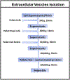
Extracellular vesicles (EVs) are cell membrane-derived compartments that regulate physiology and pathology in the body. Naturally secreted EVs have been well studied in their biogenesis and have been exploited in targeted drug delivery. Due to the limitations on production of EVs, nitrogen cavitation has been utilized to efficiently generate EV-like drug delivery systems used in treating inflammatory disorders. In this short review, we will discuss the production and purification of EVs, and we will summarize what technologies are needed to improve their production for translation. We describe the drug-loading processes in EVs and their applications as drug delivery systems for inflammatory therapies, focusing on a new type of EVs made from neutrophil membrane using nitrogen cavitation.
Keywords: drug delivery; extracellular vesicles; inflamed endothelium; neutrophils.
Figures


Similar articles
-
High yield, scalable and remotely drug-loaded neutrophil-derived extracellular vesicles (EVs) for anti-inflammation therapy.Biomaterials. 2017 Aug;135:62-73. doi: 10.1016/j.biomaterials.2017.05.003. Epub 2017 May 3. Biomaterials. 2017. PMID: 28494264 Free PMC article.
-
Extracellular vesicles for drug delivery.Adv Drug Deliv Rev. 2016 Nov 15;106(Pt A):148-156. doi: 10.1016/j.addr.2016.02.006. Epub 2016 Feb 27. Adv Drug Deliv Rev. 2016. PMID: 26928656 Review.
-
Generation, purification and engineering of extracellular vesicles and their biomedical applications.Methods. 2020 May 1;177:114-125. doi: 10.1016/j.ymeth.2019.11.012. Epub 2019 Nov 30. Methods. 2020. PMID: 31790730 Free PMC article. Review.
-
Extracellular blebs: Artificially-induced extracellular vesicles for facile production and clinical translation.Methods. 2020 May 1;177:135-145. doi: 10.1016/j.ymeth.2019.11.007. Epub 2019 Nov 14. Methods. 2020. PMID: 31734187 Review.
-
Membrane Derived Vesicles as Biomimetic Carriers for Targeted Drug Delivery System.Curr Top Med Chem. 2020;20(27):2472-2492. doi: 10.2174/1568026620666200922113054. Curr Top Med Chem. 2020. PMID: 32962615 Review.
Cited by
-
RGD-expressed bacterial membrane-derived nanovesicles enhance cancer therapy via multiple tumorous targeting.Theranostics. 2021 Jan 15;11(7):3301-3316. doi: 10.7150/thno.51988. eCollection 2021. Theranostics. 2021. PMID: 33537088 Free PMC article.
-
Human neutrophil membrane-derived nanovesicles as a drug delivery platform for improved therapy of infectious diseases.Acta Biomater. 2021 Mar 15;123:354-363. doi: 10.1016/j.actbio.2021.01.020. Epub 2021 Jan 18. Acta Biomater. 2021. PMID: 33476827 Free PMC article.
-
Liposomal Formulations Enhance the Anti-Inflammatory Effect of Eicosapentaenoic Acid in HL60 Cells.Pharmaceutics. 2022 Feb 26;14(3):520. doi: 10.3390/pharmaceutics14030520. Pharmaceutics. 2022. PMID: 35335896 Free PMC article.
-
Engineering bacterial membrane nanovesicles for improved therapies in infectious diseases and cancer.Adv Drug Deliv Rev. 2022 Jul;186:114340. doi: 10.1016/j.addr.2022.114340. Epub 2022 May 13. Adv Drug Deliv Rev. 2022. PMID: 35569561 Free PMC article. Review.
-
Neutrophil Membrane-Derived Nanovesicles Alleviate Inflammation To Protect Mouse Brain Injury from Ischemic Stroke.ACS Nano. 2019 Feb 26;13(2):1272-1283. doi: 10.1021/acsnano.8b06572. Epub 2019 Jan 28. ACS Nano. 2019. PMID: 30673266 Free PMC article.
References
-
- Duncan R, Gaspar R. Nanomedicine(s) under the microscope. Mol Pharm. 2011;8(6):2101–41. - PubMed
Publication types
MeSH terms
Substances
Grants and funding
LinkOut - more resources
Full Text Sources
Other Literature Sources

