

Quantitative axial myology in two constricting snakes: Lampropeltis holbrooki and Pantherophis obsoletus
- PMID: 29484639
- PMCID: PMC5979636
- DOI: 10.1111/joa.12799
Quantitative axial myology in two constricting snakes: Lampropeltis holbrooki and Pantherophis obsoletus
Abstract
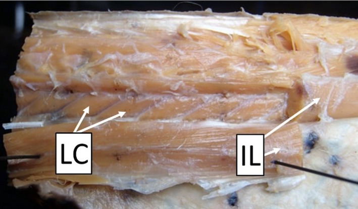
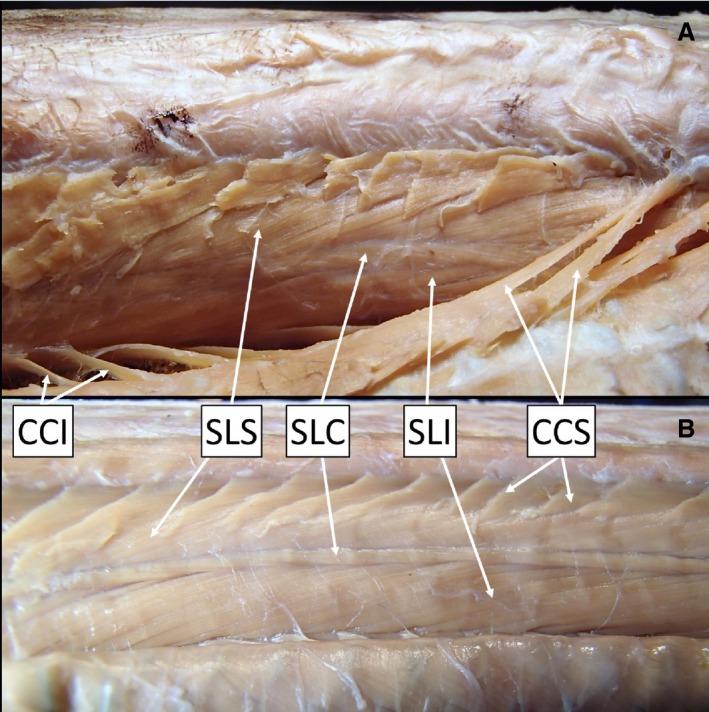
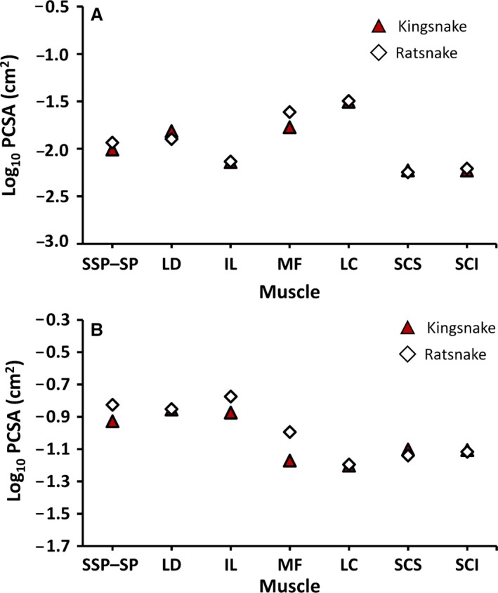
A snake's body represents an extreme degree of elongation with immense muscle complexity. Snakes have approximately 25 different muscles on each side of the body at each vertebra. These muscles serially repeat, overlap, interconnect, and rarely insert parallel to the vertebral column. The angled muscles mean that simple measurements of anatomical cross-sectional area (ACSA, perpendicular to the long-axis of the body) serve only as proxies for the primary determinant of muscle force, physiological cross-sectional area (PCSA, area perpendicular to the muscle fibers). Here, I describe and quantify the musculature of two intraguild constrictors: kingsnakes (Lampropeltis holbrooki) and ratsnakes (Pantherophis obsoletus) whose predation performance varies considerably. Kingsnakes can produce significantly higher constriction pressures compared with ratsnakes of similar size. In both snakes, I provide qualitative descriptions, detail previously undescribed complexity, identify a new lateral muscle, and provide some of the first quantitative measures of individual muscle and whole-body PCSA. Furthermore, I compare measurements of ACSA with measurements of PCSA. There was no significant difference in PCSA of muscles between kingsnakes and ratsnakes. There is, however, a strong relationship between ACSA and PCSA measurements. I could not identify a significant difference in musculature between kingsnakes and ratsnakes that explains their different levels of constriction performance. Unmeasured components of muscle function, such as endurance and force production, might account for differences in performance between two species with similar muscle structure.
Keywords: Lampropeltis; Pantherophis; anatomy; cross-sectional area; muscle; snake.
© 2018 Anatomical Society.
Figures


Similar articles
-
The king of snakes: performance and morphology of intraguild predators (Lampropeltis) and their prey (Pantherophis).J Exp Biol. 2017 Mar 15;220(Pt 6):1154-1161. doi: 10.1242/jeb.147082. J Exp Biol. 2017. PMID: 28298469
-
The scaling of terrestrial striking performance in western ratsnakes (Pantherophis obsoletus).J Exp Zool A Ecol Integr Physiol. 2020 Feb;333(2):96-103. doi: 10.1002/jez.2328. Epub 2019 Oct 17. J Exp Zool A Ecol Integr Physiol. 2020. PMID: 31625282
-
The scaling of bite force and constriction pressure in kingsnakes (Lampropeltis getula): Proximate determinants and correlated performance.Integr Zool. 2017 Mar;12(2):121-131. doi: 10.1111/1749-4877.12216. Integr Zool. 2017. PMID: 27265597
-
Considering admixture when producing draft genomes: an example in North American ratsnakes (Pantherophis alleghaniensis/Pantherophis obsoletus).G3 (Bethesda). 2023 Aug 9;13(8):jkad113. doi: 10.1093/g3journal/jkad113. G3 (Bethesda). 2023. PMID: 37228097 Free PMC article.
-
Review of the methods used for calculating physiological cross-sectional area (PCSA) for ecological questions.J Morphol. 2020 Jul;281(7):778-789. doi: 10.1002/jmor.21139. Epub 2020 May 6. J Morphol. 2020. PMID: 32374505 Review.
Cited by
-
Moving beyond the surface: Comparative head and neck myology of threadsnakes (Epictinae, Leptotyphlopidae, Serpentes), with comments on the 'scolecophidian' muscular system.PLoS One. 2019 Jul 18;14(7):e0219661. doi: 10.1371/journal.pone.0219661. eCollection 2019. PLoS One. 2019. PMID: 31318886 Free PMC article.
-
The axial anatomy of monitor lizards (Varanidae).J Anat. 2018 Nov;233(5):636-643. doi: 10.1111/joa.12872. Epub 2018 Aug 6. J Anat. 2018. PMID: 30079494 Free PMC article.
-
Corn Snakes Show Consistent Sarcomere Length Ranges Across Muscle Groups and Ontogeny.Integr Org Biol. 2022 Sep 8;4(1):obac040. doi: 10.1093/iob/obac040. eCollection 2022. Integr Org Biol. 2022. PMID: 36158732 Free PMC article.
-
Evolutionary convergence of muscle architecture in relation to locomotor ecology in snakes.J Anat. 2023 May;242(5):862-871. doi: 10.1111/joa.13823. Epub 2023 Feb 2. J Anat. 2023. PMID: 36732067 Free PMC article.
-
A Biomimetic Method to Replicate the Natural Fluid Movements of Swimming Snakes to Design Aquatic Robots.Biomimetics (Basel). 2022 Dec 3;7(4):223. doi: 10.3390/biomimetics7040223. Biomimetics (Basel). 2022. PMID: 36546923 Free PMC article.
References
-
- Alexander RM, Vernon A (1975) The dimensions of the knee and ankle muscles and the forces they exert. J Hum Mov Stud 1, 115–123.
-
- Auffenberg W (1958) The trunk musculature of Sanzinia and its bearing on certain aspects of the myological evolution in snakes. Mus Comp Zool 82, 1–12.
-
- Auffenberg W (1966) The vertebral musculature of Chersydrus (Serpentes). Q J Florida Acad Sci 29, 155–162.
-
- Cundall D (1982) Functional morphology In: Snakes: Ecology and Evolutionary Biology (eds Seigel RA, Collins JT, Novak SS.), pp. 106–140. New York: McGraw‐Hill.
-
- Cundall D, Greene HW (2000) Feeding in snakes In: Feeding: Form, Function, and Evolution in Tetrapod Vertebrates (ed. Schwenk K.), pp. 293–333. San Diego, California: Academic Press.
Publication types
MeSH terms
LinkOut - more resources
Full Text Sources
Other Literature Sources
