. 2019 Feb 15;527(3):625-639.
doi: 10.1002/cne.24419. Epub 2018 Apr 26.

Architectonic features and relative locations of primary sensory and related areas of neocortex in mouse lemurs

Affiliations

Architectonic features and relative locations of primary sensory and related areas of neocortex in mouse lemurs

Mansi P Saraf et al. J Comp Neurol. .

Abstract

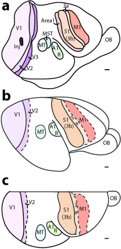
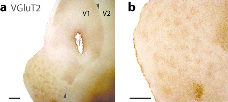
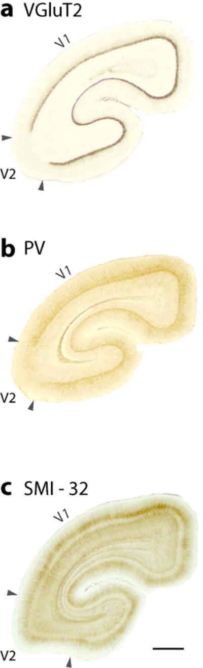
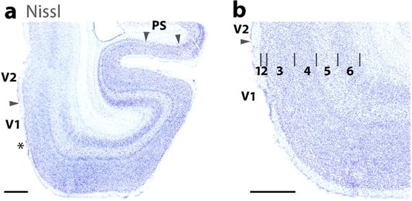
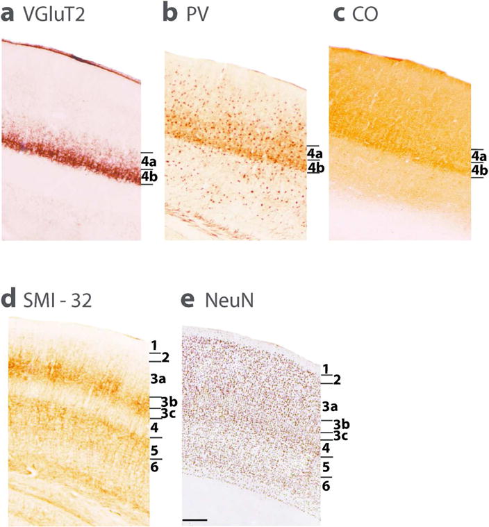
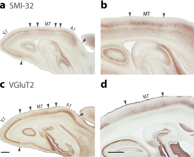
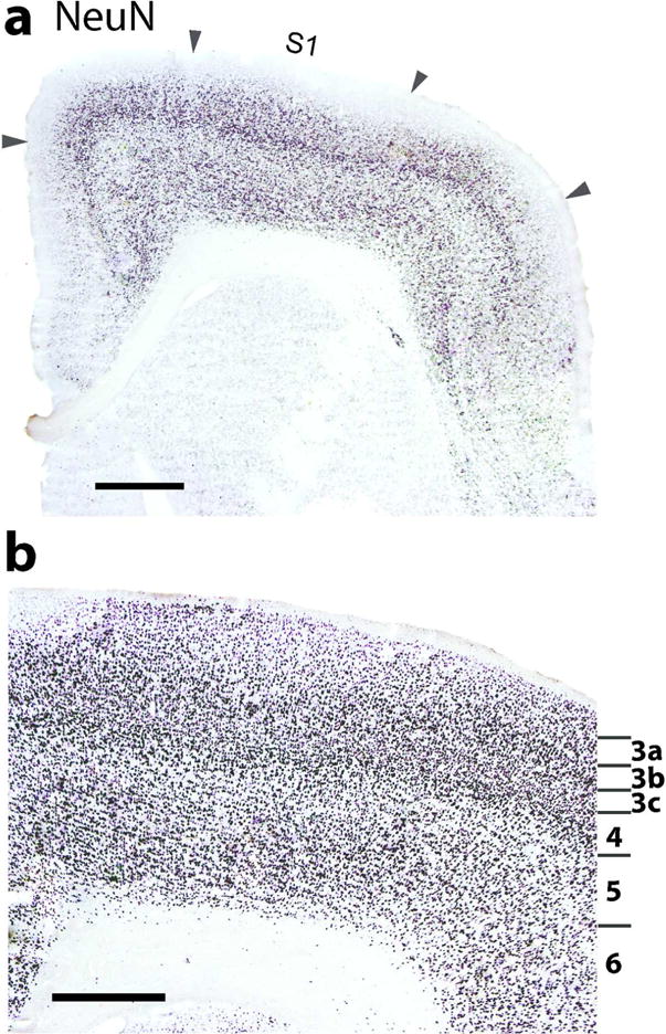
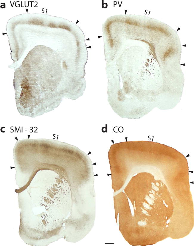
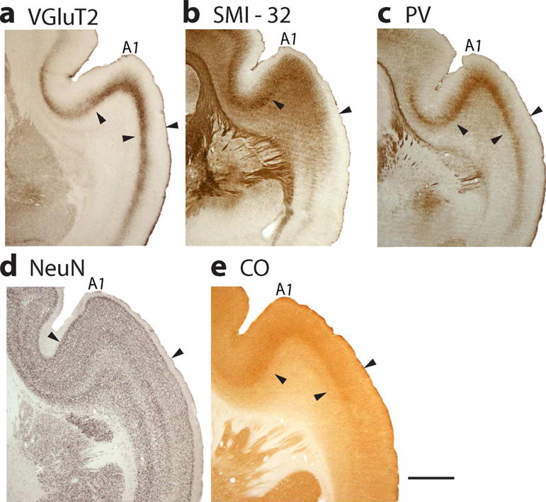
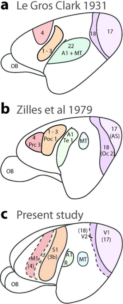
Mouse lemurs are the smallest of the living primates, and are members of the understudied radiation of strepsirrhine lemurs of Madagascar. They are thought to closely resemble the ancestral primates that gave rise to present day primates. Here we have used multiple histological and immunochemical methods to identify and characterize sensory areas of neocortex in four brains of adult lemurs obtained from a licensed breeding colony. We describe the laminar features for the primary visual area (V1), the secondary visual area (V2), the middle temporal visual area (MT) and area prostriata, somatosensory areas S1(3b), 3a, and area 1, the primary motor cortex (M1), and the primary auditory cortex (A1). V1 has "blobs" with "nonblob" surrounds, providing further evidence that this type of modular organization might have evolved early in the primate lineage to be retained in all extant primates. The laminar organization of V1 further supports the view that sublayers of layer 3 of primates have been commonly misidentified as sublayers of layer 4. S1 (area 3b) is proportionately wider than the elongated area observed in anthropoid primates, and has disruptions that may distinguish representations of the hand, face, teeth, and tongue. Primary auditory cortex is located in the upper temporal cortex and may include a rostral area, R, in addition to A1. The resulting architectonic maps of cortical areas in mouse lemurs can usefully guide future studies of cortical connectivity and function.

Keywords: RRID_ AB_177621; RRID_ AB_2313581; RRID_AB_2313581; RRID_AB_2564642; RRID_AB_477329; auditory cortex; primates; prosimian evolution; somatosensory cortex; visual cortex.


Conflict of interest statement

Conflict of Interest

The authors declare no conflict of interest.

Figures

Figure 1
Figure 1
The locations of sensory and motor areas of neocortex in mouse lemur as defined by six different architectonic procedures. (a) A surface view of cortex based on brain sections cut parallel to the surface of flattened cortex; (b) a lateral view based on coronal sections; and (c) a dorsal view based on sagittal sections. V1, V2 and V3, visual areas 1–3; MST and MT, middle temporal visual areas and medial superior temporal area; R and A1, auditory area 1 and the rostral auditory area; S1, 3b, 3a and 1;somatosensory areas S1(3b), 3a and 1; M1, primary motor cortex; OB, olfactory bulb. Inj, a damaged region due to an injection of a tracer. Scale bar = 1 mm.
Figure 2
Figure 2
Caudal parts of brain sections cut parallel to flattened cortex and processed for vesicular glutamate transporter 2 (VGluT2). (a) Lower magnification shows a clear border between V1 and V2. The darker parts along upper border reflect the lateral geniculate nucleus (LGN) projection terminations in layer 4, while the pattern of dots in the lower part of the section reflect the layer 3 that are characterized by LGN inputs. (b) A higher magnification of the part of the section with blobs. Scale bar = 1 mm.
Figure 3
Figure 3
Coronal brain sections from occipital cortex showing the architectonic features of V1 (area17) and V2 (area18). The sections have been processed for, (a) vesicular glutamate transporter 2 (VGlut2), (b) Parvalbumin (PV) and (c) SMI-32. Scale bar = 1 mm.
Figure 4
Figure 4
The laminar characteristics of areas 17 (V1), 18 (V2), and Prostriata (PS) in a horizontal brain section stained for Nissl substance. (a) A lower magnification photo of ventromedial V1 (17) and V2 (18). Arrows mark the 17–18 boundary and the 17/prostriata boundary (PS). A transition zone (marked by asterisk) is apparent in area 17 near the area 18 boundary where some of the laminar features of area 17 are less apparent. Prostriata (arrow heads) is also prominent in the section. (b) A higher magnification of the laminar pattern of area 17. Note the sublaminar pattern in layer 4. Scale bar = 1 mm.
Figure 5
Figure 5
Laminar characteristics of area 17 (V1) in a coronal brain sections processed for VGluT2 (a), Parvalbumin (b), Cytochrome oxidase (c), SMI-32 (d) and NeuN (e). A, B, and C, show two subdivsions of layer 4. SMI-32 (d) stains pyramidal neurons in layer 3 and 5. In NeuN preparations (e), the darkly stained neuron cell bodies mark layers 4,6,3 and 2 as darker than layers 5 and 3 c.
Figure 6
Figure 6
Coronal brain sections through occipital temporal cortex processed for SMI-32 (a, b) or VGluT2 (c, d). (a) MT is seen in the section processed for SMI-32, between area 17 and the auditory cortex. SMI-32 marks pyramidal neurons of layers 3 and 5. (b) Higher magnification of MT (c) The presumptive area MT is characterized by the expression of VGluT2 in layer 4. Area 17 (V1) is also defined by dense VGluT2 expression in layer 4 along with auditory cortex (A1). (d) Higher magnification of the part of the section showing MT. Scale bar in a = 1 mm, in b = 0.5 mm.
Figure 7
Figure 7
A coronal section of S1 cortex processed for neuron cell bodies with NeuN. (a) At a lower magnification, S1 (area 3b) appears to be patchy (arrowheads) due to disjunctions in the S1 representation (likely representing foot and hand). (b) A view of S1 at a higher magnification marking layers 3 through 6. Layers 3 and 4 are densely packed with neurons. Also note the sublaminar pattern in layer 3. Scale bar in a = 1 mm, in b = 0.5 mm.
Figure 8
Figure 8
S1 in especially more frontal sections appears to be patchy, due to disjunctions in the somatotopy. (a) Coronal section through frontal cortex processed for VGluT2. Dense patches of label (marked by arrowheads) in layer 4 and extending into layer 3, characterize parts of S1 (3b) likely representing the foot and hand. A lateral patch (arrow heads) may correspond to mouth representation. (b) PV is expressed densely in layer 4 and somewhat in layer 3 of S1. (c) Section processed for SMI-32 has dense patches of labeled neurons, mainly in layer 3 of S1, but also in layer 5. The dorsolateral patches likely represent the foot and hand of S1, while a medial patch may represent the tail and hand limb. A most lateral patch could be S1 teeth or tongue cortex. (d) The S1 patches are also apparent in a sections processed for CO. Scale bar = 1 mm.
Figure 9
Figure 9
Primary Motor cortex, M1, is located rostromedial to S1 and 3a (Figure 1). Coronal sections processed for Nissl (A, B) and VGluT2 (c, d) show the relative locations and architectonic features of these areas as shown at higher magnification (b, d). In Nissl preparations, layer 4 of small cells is well developed in area 3b, reduced in dysgranular 3a, and greatly reduced or absent in agranular M1. In VGluT2 preparations, layer 4 of area 3a is less darkly stained, and the layer 3–4 region of M1 is lightly stained Arrow heads mark boundaries. Scale bar in a and c = 1 mm, in b and d = 0.5 mm.
Figure 10
Figure 10
Primary auditory cortex, A1, is located in the dorsal temporal lobe where it extends into the lateral sulcus. Features that characterize primary sensory cortex in A1 are shown in coronal sections processed for (a) VGluT2, (b) SMI-32, (c) PV, (d) NeuN, and (e) CO. Scale bar = 1 mm.
Figure 11
Figure 11
Comparisons of illustrated summary diagrams from earlier architecture studies in mouse lemurs (a and b) and present results (c). The earlier studies included more architectonics fields but the redrawn diagrams shown here only include proposed sensory and motor fields relevant to present results. Scale bar = 1 mm.

Similar articles

Cited by

References

    1. Allman JM, Kaas JH. Representation of the visual field in striate and adjoining cortex of the owl monkey (Aotus trivirgatus) Brain Research. 1971;35(1):89–106. - PubMed
    1. Allman JM, Kaas JH, Lane RH. The middle temporal visual area (MT) in the bush baby, Galago senegalensis. Brain Res. 1973;57:197–202. - PubMed
    1. Allman J. Variations in visual cortex organization in primates. Neurobiology of Neocortex. 1988:249–40.
    1. Azevedo FA, Carvalho LR, Grinberg LT, Farfel JM, Ferretti RE, Leite RE, Herculano Houzel S. Equal numbers of neuronal and nonneuronal cells make the human brain an isometrically scaled up primate brain. Journal of Comparative Neurology. 2009;513(5):532–541. - PubMed
    1. Balaram P, Kaas JH. Towards a unified scheme of cortical lamination for primary visual cortex across primates: insights from NeuN and VGLUT2 immunoreactivity. Frontiers in neuroanatomy. 2014;8:81. - PMC - PubMed

Publication types

MeSH terms

Substances

LinkOut - more resources