

Iron deficiency impairs contractility of human cardiomyocytes through decreased mitochondrial function
- PMID: 29484788
- PMCID: PMC5993224
- DOI: 10.1002/ejhf.1154
Iron deficiency impairs contractility of human cardiomyocytes through decreased mitochondrial function
Abstract
Aims: Iron deficiency is common in patients with heart failure and associated with a poor cardiac function and higher mortality. How iron deficiency impairs cardiac function on a cellular level in the human setting is unknown. This study aims to determine the direct effects of iron deficiency and iron repletion on human cardiomyocytes.
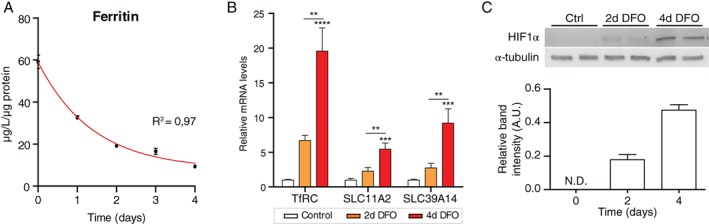
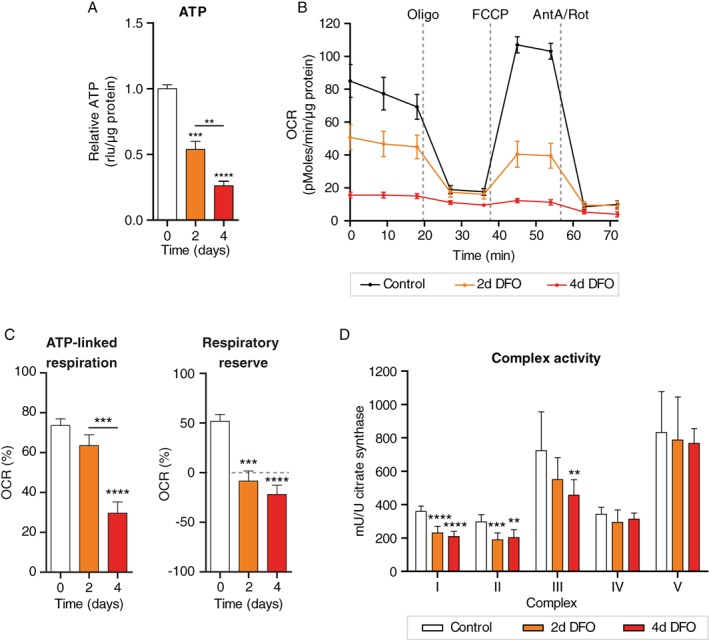
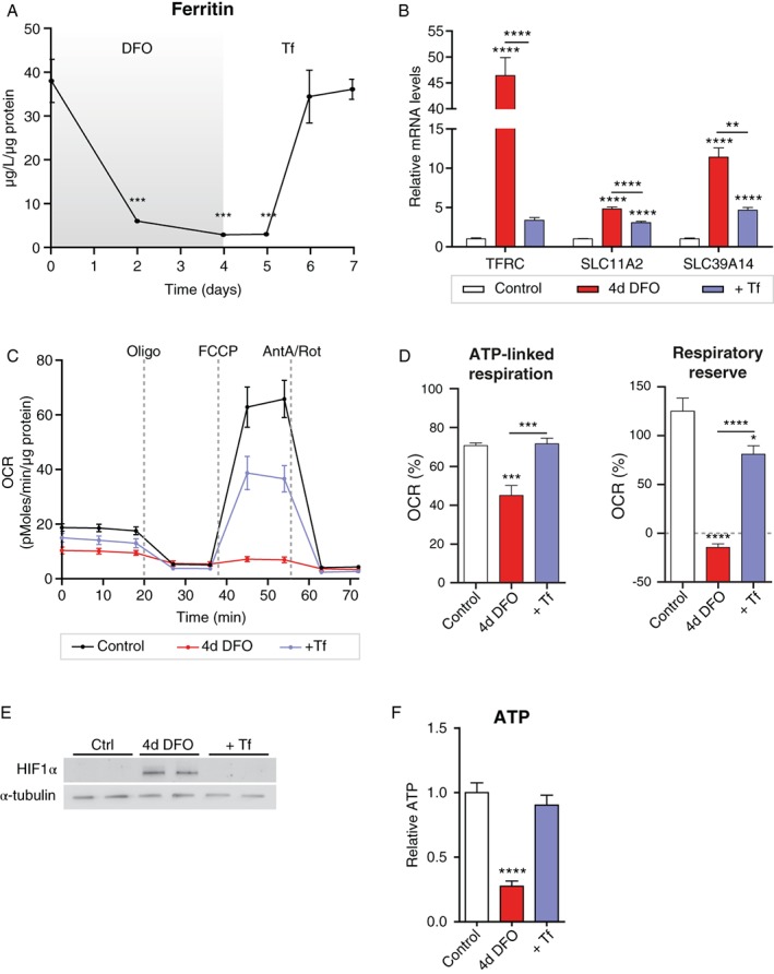
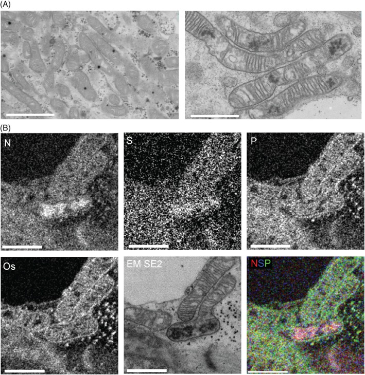
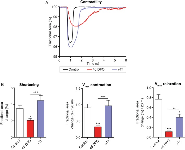
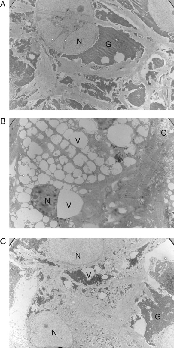
Methods and results: Human embryonic stem cell-derived cardiomyocytes were depleted of iron by incubation with the iron chelator deferoxamine (DFO). Mitochondrial respiration was determined by Seahorse Mito Stress test, and contractility was directly quantified using video analyses according to the BASiC method. The activity of the mitochondrial respiratory chain complexes was examined using spectrophotometric enzyme assays. Four days of iron depletion resulted in an 84% decrease in ferritin (P < 0.0001) and significantly increased gene expression of transferrin receptor 1 and divalent metal transporter 1 (both P < 0.001). Mitochondrial function was reduced in iron-deficient cardiomyocytes, in particular ATP-linked respiration and respiratory reserve were impaired (both P < 0.0001). Iron depletion affected mitochondrial function through reduced activity of the iron-sulfur cluster containing complexes I, II and III, but not complexes IV and V. Iron deficiency reduced cellular ATP levels by 74% (P < 0.0001) and reduced contractile force by 43% (P < 0.05). The maximum velocities during both systole and diastole were reduced by 64% and 85%, respectively (both P < 0.001). Supplementation of transferrin-bound iron recovered functional and morphological abnormalities within 3 days.
Conclusion: Iron deficiency directly affects human cardiomyocyte function, impairing mitochondrial respiration, and reducing contractility and relaxation. Restoration of intracellular iron levels can reverse these effects.
Keywords: Contractility; Heart failure; Human cardiomyocytes; Iron deficiency.
© 2018 The Authors. European Journal of Heart Failure published by John Wiley & Sons Ltd on behalf of European Society of Cardiology.
Figures
Comment in
-
Cardiac iron deficiency-how to refuel the engine out of fuel.Eur J Heart Fail. 2018 May;20(5):920-922. doi: 10.1002/ejhf.1174. Epub 2018 Mar 1. Eur J Heart Fail. 2018. PMID: 29493065 No abstract available.
References
-
- Jankowska EA, Rozentryt P, Witkowska A, Nowak J, Hartmann O, Ponikowska B, Borodulin‐Nadzieja L, Banasiak W, Polonski L, Filippatos G, McMurray JJ, Anker SD, Ponikowski P. Iron deficiency: an ominous sign in patients with systolic chronic heart failure. Eur Heart J 2010;31:1872–1880. - PubMed
-
- van Veldhuisen DJ, Anker SD, Ponikowski P, Macdougall IC. Anemia and iron deficiency in heart failure: mechanisms and therapeutic approaches. Nat Rev Cardiol 2011;8:485–493. - PubMed
-
- Ponikowski P, Voors AA, Anker SD, Bueno H, Cleland JG, Coats AJ, Falk V, González‐Juanatey JR, Harjola VP, Jankowska EA, Jessup M, Linde C, Nihoyannopoulos P, Parissis JT, Pieske B, Riley JP, Rosano GM, Ruilope LM, Ruschitzka F, Rutten FH, van der Meer P. 2016 ESC Guidelines for the diagnosis and treatment of acute and chronic heart failure: The Task Force for the diagnosis and treatment of acute and chronic heart failure of the European Society of Cardiology (ESC). Developed with the special contribution of the Heart Failure Association (HFA) of the ESC. Eur J Heart Fail 2016;18:891–975. - PubMed
-
- Comín‐Colet J, Enjuanes C, González G, Torrens A, Cladellas M, Meroño O, Ribas N, Ruiz S, Gómez M, Verdú JM, Bruguera J. Iron deficiency is a key determinant of health‐related quality of life in patients with chronic heart failure regardless of anaemia status. Eur J Heart Fail 2013;15:1164–1172. - PMC - PubMed
Publication types
MeSH terms
LinkOut - more resources
Full Text Sources
Other Literature Sources
Medical
