Biomechanical Analysis of a Novel Intercalary Prosthesis for Humeral Diaphyseal Segmental Defect Reconstruction
- PMID: 29484857
- PMCID: PMC6594488
- DOI: 10.1111/os.12368
Biomechanical Analysis of a Novel Intercalary Prosthesis for Humeral Diaphyseal Segmental Defect Reconstruction
Abstract
Objective: To study the biomechanical properties of a novel modular intercalary prosthesis for humeral diaphyseal segmental defect reconstruction, to establish valid finite element humerus and prosthesis models, and to analyze the biomechanical differences in modular intercalary prostheses with or without plate fixation.
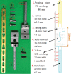
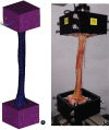
Methods: Three groups were set up to compare the performance of the prosthesis: intact humerus, humerus-prosthesis and humerus-prosthesis-plate. The models of the three groups were transferred to finite element software. Boundary conditions, material properties, and mesh generation were set up for both the prosthesis and the humerus. In addition, 100 N or 2 N.m torsion was loaded to the elbow joint surface with the glenohumeral joint surface fixed. Humeral finite element models were established according to CT scans of the cadaveric bone; reverse engineering software Geomagic was used in this procedure. Components of prosthetic models were established using 3-D modeling software Solidworks. To verify the finite element models, the in vitro tests were simulated using a mechanical testing machine (Bionix; MTS Systems Corporation, USA). Starting with a 50 N preload, the specimen was subjected to 5 times tensile (300 N) and torsional (5 N.m) strength; interval time was 30 min to allow full recovery for the next specimen load. Axial tensile and torsional loads were applied to the elbow joint surface to simulate lifting heavy objects or twisting something, with the glenohumeral joint surface fixed.
Results: Stress distribution on the humerus did not change its tendency notably after reconstruction by intercalary prosthesis whether with or without a plate. The special design which included a plate and prosthesis effectively diminished stress on the stem where aseptic loosening often takes place. Stress distribution major concentrate upon two stems without plate addition, maximum stress on proximal and distal stem respectively diminish 27.37% and 13.23% under tension, 10.66% and 11.16% under torsion after plate allied.
Conclusion: The novel intercalary prosthesis has excellent ability to reconstruct humeral diaphyseal defects. The accessory fixation system, which included a plate and prosthesis, improved the rigidity of anti-tension and anti-torsion, and diminished the risk of prosthetic loosening and dislocation. A finite element analysis is a kind of convenient and practicable method to be used as the confirmation of experimental biomechanics study.
Keywords: Biomechanics; Bone tumor; Finite element analysis; Intercalary prosthesis.
© 2018 Chinese Orthopaedic Association and John Wiley & Sons Australia, Ltd.
Figures


References
-
- Wedin R, Hansen BH, Laitinen M, et al Complications and survival after surgical treatment of 214 metastatic lesions of the humerus. J Shoulder Elbow Surg, 2012, 21: 1049–1055. - PubMed
-
- Harrington KD. Orthopaedic management of extremity and pelvic lesions. Clin Orthop Relat Res, 1995, 312: 136–147. - PubMed
-
- Rock MG. Metastatic lesions of the humerus and the upper extremity. Instr Course Lect, 1992, 41: 114–115. - PubMed
-
- Chapman JR, Henley MB, Aqel J, Benca PJ. Randomized prospective study of humeral shaft fracture fixation: intramedullary nails versus plates. J Orthop Trauma, 2000, 14: 162–166. - PubMed
-
- Farragos AF, Schemitsch EH, McKee MD. Complications of intramedullary nailing for fractures of the humeral shaft: a review. J Orthop Trauma, 1999, 13: 258–267. - PubMed
MeSH terms
LinkOut - more resources
Full Text Sources
Other Literature Sources

