. 2019 Jul;39(7):1355-1368.
doi: 10.1177/0271678X18760974. Epub 2018 Feb 27.

Delayed clearance of cerebrospinal fluid tracer from entorhinal cortex in idiopathic normal pressure hydrocephalus: A glymphatic magnetic resonance imaging study

Affiliations

Delayed clearance of cerebrospinal fluid tracer from entorhinal cortex in idiopathic normal pressure hydrocephalus: A glymphatic magnetic resonance imaging study

Per K Eide et al. J Cereb Blood Flow Metab. 2019 Jul.

Abstract

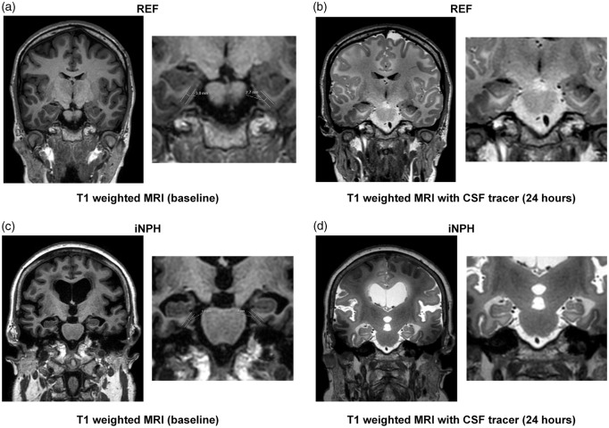
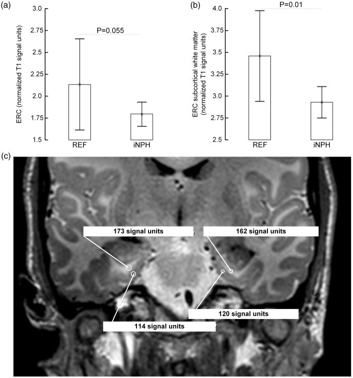
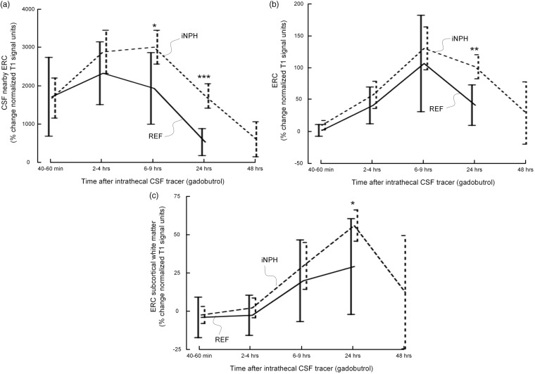
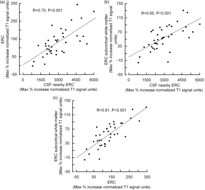
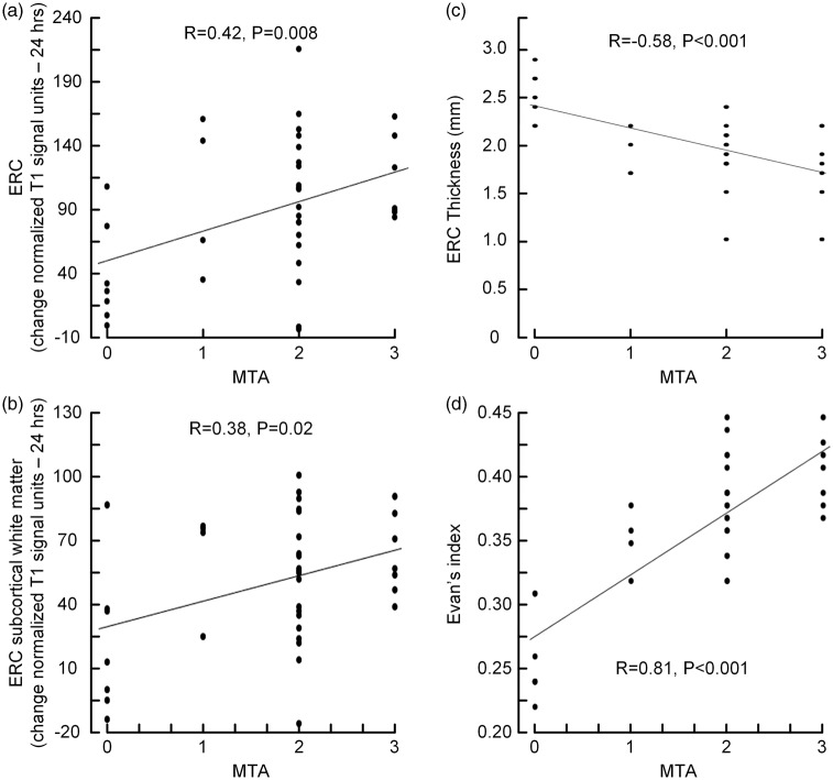
The glymphatic system plays a key role for clearance of waste solutes from the rodent brain. We recently found evidence of glymphatic circulation in the human brain when using magnetic resonance imaging (MRI) contrast agent as cerebrospinal fluid (CSF) tracer in conjunction with multiple MRI acquisitions (gMRI). The present study explored the hypothesis that reduced glymphatic clearance in entorhinal cortex (ERC) may be instrumental in idiopathic normal pressure hydrocephalus (iNPH) dementia. gMRI acquisitions were obtained over a 24-48 h time span in cognitively affected iNPH patients and non-cognitively affected patients with suspected CSF leaks. The CSF tracer enrichment was determined as changes in normalized MRI T1 signal units. The study included 30 patients with iNPH and 8 individuals with suspected CSF leaks (i.e. reference individuals). Compared to reference individuals, iNPH patients presented with higher medial temporal lobe atrophy score and Evan's index and inferior ERC thickness. We found delayed clearance of the intrathecal CSF tracer gadobutrol from CSF, the ERC and adjacent white matter, suggesting impaired glymphatic circulation. Reduced clearance and accumulation of toxic waste product such as amyloid-β may be a mechanism behind dementia in iNPH. Glymphatic MRI (gMRI) may become a tool for assessment of early dementia.

Keywords: Idiopathic normal pressure hydrocephalus; cerebrospinal fluid tracer; dementia; entorhinal cortex; glymphatic circulation.


Figures

Figure 1.
Figure 1.
In an REF individual (a, b) and an iNPH patient (c, d) T1-weighted MRI in coronal sections at ERC level at baseline (before contrast agent administration) (a and c) and after 24 h (b and d) illustrate time-dependent enrichment of CSF by increased T1 image intensity at the image greyscale. Ventricular reflux of contrast agent is a typical feature of iNPH (d) as described previously.
Figure 2.
Figure 2.
The normalized T1 signal units before intrathecal administration of gadobutrol in REF and iNPH patients within (a) ERC, and (b) subcortical white matter of ERC. Differences between REF and iNPH individuals are indicated for (a) ERC and (b) ERC subcortical white matter, and may be attributed to increased brain water content in iNPH. In (c) is shown how ROIs for measurement of MRI T1 signal units within the ERC and subcortical white matter of ERC were manually fitted in an REF subject. All T1 signal unit measurements were normalized against a reference value retrieved from the superior sagittal sinus, which does not enhance with intrathecal contrast agent.
Figure 3.
Figure 3.
Trend plots of time-dependent CSF tracer (gadobutrol) uptake in REF (continuous lines) and iNPH (dotted lines) patients revealed as percentage change in normalized T1 signal unit from baseline at three locations: (a) CSF nearby ERC, (b) ERC and (c) subcortical white matter of ERC. Error bars refer to 95% CI. Differences between groups at individual time points were determined by mixed model analysis (*p < 0.05, **p < 0.01, ***p < 0.001). In iNPH cases, the CSF tracer enhancement was significantly higher at all locations at 24 h, indicative of delayed tracer clearance. Peak enhancement in ERC (b) precedes peak enhancement in ERC subcortical white matter (c), demonstrating a centripetal pattern of brain enrichment by CSF tracer.
Figure 4.
Figure 4.
CSF tracer uptake within ERC is a function of tracer enhancement within CSF nearby ERC. There was a highly significant positive correlation between CSF tracer availability within the CSF space nearby ERC and (a) ERC and (b) subcortical white matter of ERC, as well as (c) between ERC and subcortical white matter of ERC. For each plot is presented the fit line and the Pearson correlation coefficient (R) with P-value.
Figure 5.
Figure 5.
Association between MTA-score and percentage increase of normalized T1 signal units (i.e. CSF tracer enrichment) at 24 h as compared to before contrast agent administration within (a) ERC, and within (b) subcortical white matter of ERC. Further, the associations between MTA-score and (c) ERC thickness and (d) Evans index are indicated. For each plot is presented the fit line and the Pearson correlation coefficient (R) with significance P-value.

Comment in

Similar articles

Cited by

References

    1. Moser MB, Rowland DC, Moser EI. Place cells, grid cells, and memory. Cold Spring Harbor Perspect Biol 2015; 7: a021808. - PMC - PubMed
    1. Pennanen C, Kivipelto M, Tuomainen S, et al. Hippocampus and entorhinal cortex in mild cognitive impairment and early AD. Neurobiol Aging 2004; 25: 303–310. - PubMed
    1. Hyman BT, Van Hoesen GW, Damasio AR, et al. Alzheimer's disease: cell-specific pathology isolates the hippocampal formation. Science 1984; 225: 1168–1170. - PubMed
    1. Thaker AA, Weinberg BD, Dillon WP, et al. Entorhinal cortex: antemortem cortical thickness and postmortem neurofibrillary tangles and amyloid pathology. Am J Neuroradiol 2017; 38: 961–965. - PMC - PubMed
    1. Iliff JJ, Wang M, Liao Y, et al. A paravascular pathway facilitates CSF flow through the brain parenchyma and the clearance of interstitial solutes, including amyloid beta. Sci Transl Med 2012; 4: 147ra11. - PMC - PubMed

LinkOut - more resources