. 2018 Feb 27:5:180006.
doi: 10.1038/sdata.2018.6.

An open repository for single-cell reconstructions of the brain forest

Affiliations

An open repository for single-cell reconstructions of the brain forest

Masood A Akram et al. Sci Data. .

Abstract

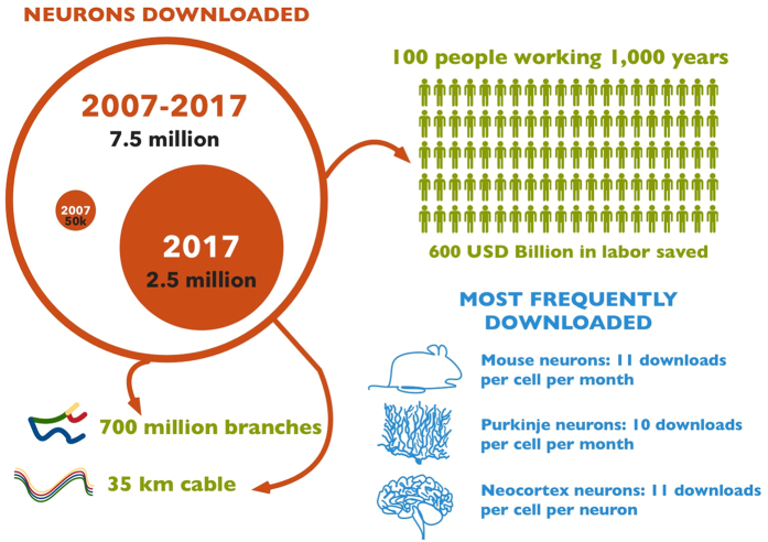
NeuroMorpho.Org was launched in 2006 to provide unhindered access to any and all digital tracings of neuronal morphology that researchers were willing to share freely upon request. Today this database is the largest public inventory of cellular reconstructions in neuroscience with a content of over 80,000 neurons and glia from a representative diversity of animal species, anatomical regions, and experimental methods. Datasets continuously contributed by hundreds of laboratories worldwide are centrally curated, converted into a common non-proprietary format, morphometrically quantified, and annotated with comprehensive metadata. Users download digital reconstructions for a variety of scientific applications including visualization, classification, analysis, and simulations. With more than 1,000 peer-reviewed publications describing data stored in or utilizing data retrieved from NeuroMorpho.Org, this ever-growing repository can already be considered a mature resource for neuroscience.


Conflict of interest statement

The authors declare no competing financial interests.

Figures

Figure 1
Figure 1. Digital reconstructions of cellular morphologies in NeuroMorpho.Org.
(a) Microglia cell from mouse spinal cord, with processes in blue and cell body in red (Data Citation 1). (b) Ganglion cell from mouse retina, with dendrites in green, axon in silver, and soma in red (Data Citation 2). (c) Pyramidal neuron from mouse neocortex, with apical dendrites in magenta, basal dendrites in green, and long projection axon in silver (Data Citation 3). (d) Pyramidal neuron from rat hippocampus, with apical dendrites in magenta, basal dendrites in green, and (incomplete) axon in silver (Data Citation 4). (e) Interneuron from mouse retina, with unspecified neurites in pink (Data Citation 5). (f) Direction sensitive mechanoreceptor from cricket peripheral nervous system, with unspecified neurites in pink (Data Citation 6). (g) Olivocerebellar neuron from rat myelencephalon, with axon in silver (Data Citation 7). (h) Pyramidal neuron from cat neocortex, with apical dendrites in magenta, basal dendrites in green, and long projection axon in silver (Data Citation 8).
Figure 2
Figure 2. Browse functionality.
(a) Summary view of selected neurons from the Browse-by-Archive pane, with links to individual entries and options for download. (b) NeuronAtlas, a free downloadable application for exploring digital reconstructions of neuronal morphologies from rat and mouse brains.
Figure 3
Figure 3. NeuroMorpho.Org’s API gives developers and data miners access to database content and information allowing integration with other neuroinformatics tools.
Complete usage documentation and definitions are available at neuromorpho.org/apiReference.html. (a) Sample of available JSON objects characterizing experimental methods. (b) Selected entries specifying the dimension of cell types. (c) Subset of a given cell’s morphometric characteristics defined as attribute-value pairs. (d) Bibliographic details for a data set from an individual publication.
Figure 4
Figure 4. Infographics summary of data download activity from the NeuroMorpho.Org portal.
Figure 5
Figure 5. Flowchart of the operational procedures of NeuroMorpho.Org from start (Literature search) to end (Main site release), highlighting literature mining (blue), morphological standardization (green), metadata annotation (pink), and data release (yellow).
Figure 6
Figure 6. Literature database and search.
(a) Retrievable information by availability status. To avoid singling out individual authors, the identifiers for declined or unanswered queries are fictional in this illustration; actual unabridged data are publicly posted online in the ‘Literature Coverage’ section of NeuroMorpho.Org. (b) Preliminary metadata annotation for all references deemed to describe digital reconstructions of neuronal morphology.

References

Data Citations

    1. Ohgomori T. 2017. NeuroMorpho.Org. NMO_66876
    1. Badea T. C. 2014. NeuroMorpho.Org. NMO_10752
    1. Chandrashekar J. 2017. NeuroMorpho.Org. NMO_51118
    1. Amaral D. G. 2006. NeuroMorpho.Org. NMO_00940
    1. Denk W. 2017. NeuroMorpho.Org. NMO_63366

References

    1. Sejnowski T. J., Koch C. & Churchland P. S. Computational neuroscience. Science (New York, N.Y.) 241, 1299–1306 (1988). - PubMed
    1. Dayan P. Computational modelling. Current opinion in neurobiology 4, 212–217 (1994). - PubMed
    1. Hines M. L. & Carnevale N. T. NEURON: a tool for neuroscientists. The Neuroscientist: a review journal bringing neurobiology, neurology and psychiatry 7, 123–135 (2001). - PubMed
    1. Senft S. L. A brief history of neuronal reconstruction. Neuroinformatics 9, 119–128 (2011). - PubMed
    1. Ascoli G. A. Mobilizing the base of neuroscience data: the case of neuronal morphologies. Nature reviews. Neuroscience 7, 318–324 (2006). - PubMed

Publication types