

Analysis of 14-3-3 isoforms expressed in photoreceptors
- PMID: 29486162
- PMCID: PMC5924652
- DOI: 10.1016/j.exer.2018.02.022
Analysis of 14-3-3 isoforms expressed in photoreceptors
Abstract
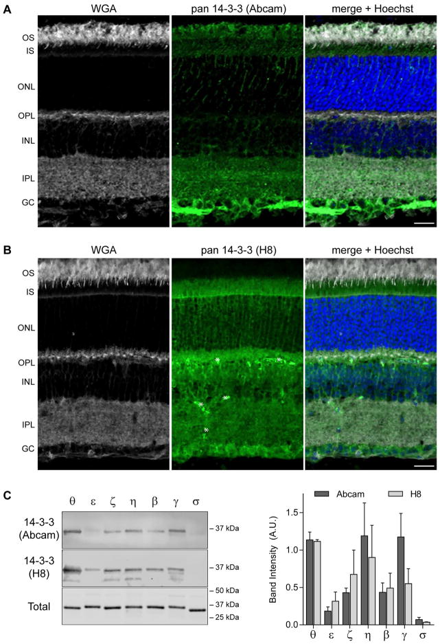
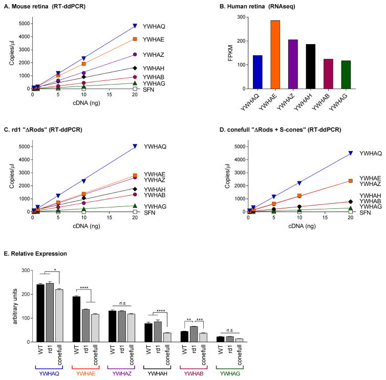
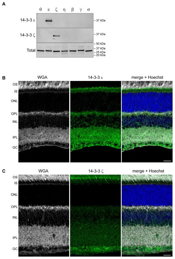
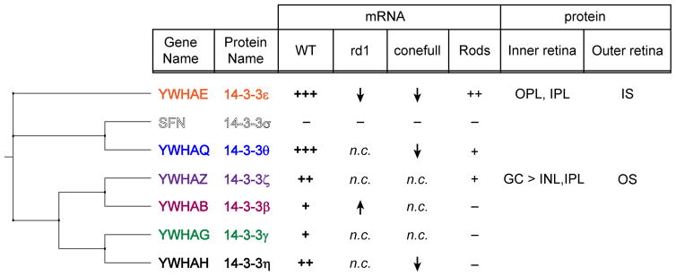
The 14-3-3 family of proteins has undergone considerable expansion in higher eukaryotes with humans and mice expressing seven isoforms (β, ε, η, γ, θ, ζ, and σ) from seven distinct genes (YWHAB, YWAHE, YWHAH, YWHAG, YWHAQ, YWHAZ, and SFN). Growing evidence indicates that while highly conserved, these isoforms are not entirely functionally redundant as they exhibit unique tissue expression profiles, subcellular localization, and biochemical functions. A key limitation in our understanding of 14-3-3 biology lies in our limited knowledge of cell-type specific 14-3-3 expression. Here we provide a characterization of 14-3-3 expression in whole retina and isolated rod photoreceptors using reverse-transcriptase digital droplet PCR. We find that all 14-3-3 genes with the exception of SFN are expressed in mouse retina with YWHAQ and YWHAE being the most highly expressed. Rod photoreceptors are enriched in YWHAE (14-3-3 ε). Immunohistochemistry revealed that 14-3-3 ε and 14-3-3 ζ exhibit unique distributions in photoreceptors with 14-3-3 ε restricted to the inner segment and 14-3-3 ζ localized to the outer segment. Our data demonstrates that, in the retina, 14-3-3 isoforms likely serve specific functions as they exhibit unique expression levels and cell-type specificity. As such, future investigations into 14-3-3 function in rod photoreceptors should be centered on 14-3-3 ε and 14-3-3 ζ, depending on the subcellular region of question.
Keywords: 14-3-3; Cone; Digital droplet PCR; Photoreceptor; Retina; YWHAE; YWHAZ; rd1.
Copyright © 2018 Elsevier Ltd. All rights reserved.
Conflict of interest statement
Figures
References
-
- Aitken A. 14-3-3 proteins: a historic overview. Semin Cancer Biol. 2006;16:162–172. - PubMed
-
- Bridges D, Moorhead GB. 14-3-3 proteins: a number of functions for a numbered protein. Sci STKE. 2005;2005:re10. - PubMed
-
- Buret L, Rebillard G, Brun E, Angebault C, Pequignot M, Lenoir M, Do-Cruzeiro M, Tournier E, Cornille K, Saleur A, Gueguen N, Reynier P, Amati-Bonneau P, Barakat A, Blanchet C, Chinnery P, Yu-Wai-Man P, Kaplan J, Roux AF, Van Camp G, Wissinger B, Boespflug-Tanguy O, Giraudet F, Puel JL, Lenaers G, Hamel C, Delprat B, Delettre C. Loss of function of in mice induces deafness and cochlear outer hair cells’ degeneration. Cell death discovery. 2016;2:16017. - PMC - PubMed
-
- Bustos DM, Iglesias AA. Intrinsic disorder is a key characteristic in partners that bind 14-3-3 proteins. Proteins. 2006;63:35–42. - PubMed
Publication types
MeSH terms
Substances
Grants and funding
LinkOut - more resources
Full Text Sources
Other Literature Sources
Research Materials
Miscellaneous
