. 2018 Feb 26;44(4):433-446.e7.
doi: 10.1016/j.devcel.2018.01.021.

Myocardial Polyploidization Creates a Barrier to Heart Regeneration in Zebrafish

Affiliations

Myocardial Polyploidization Creates a Barrier to Heart Regeneration in Zebrafish

Juan Manuel González-Rosa et al. Dev Cell. .

Abstract

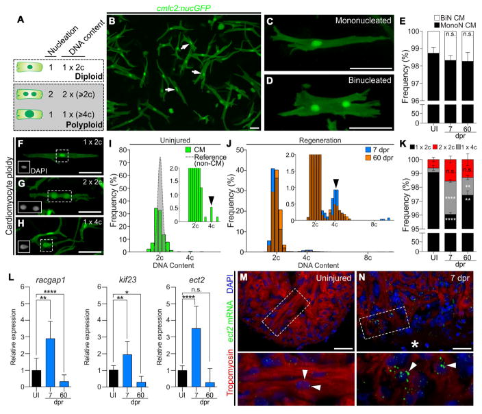
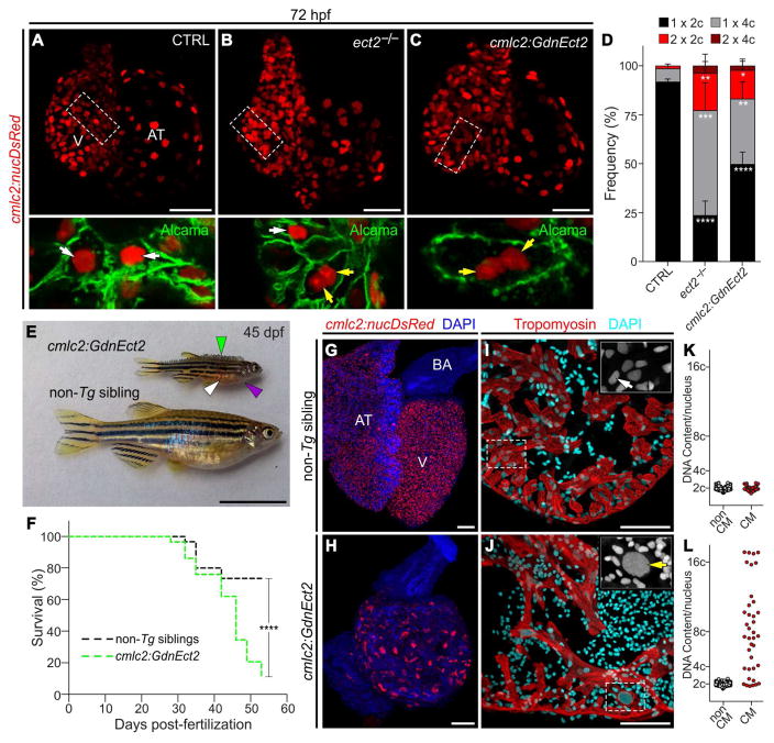
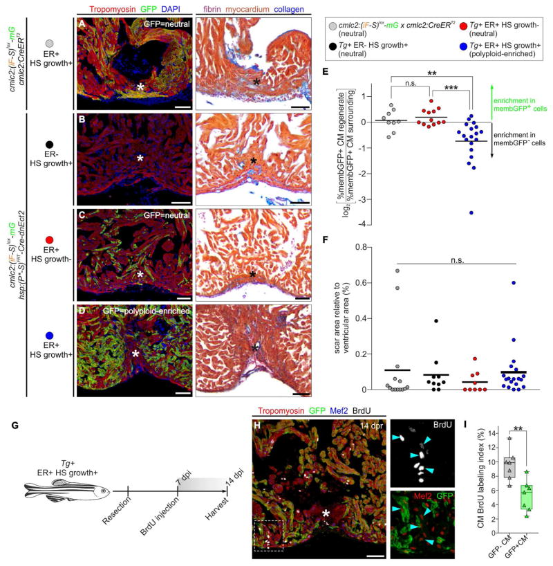
Correlative evidence suggests that polyploidization of heart muscle, which occurs naturally in post-natal mammals, creates a barrier to heart regeneration. Here, we move beyond a correlation by demonstrating that experimental polyploidization of zebrafish cardiomyocytes is sufficient to suppress their proliferative potential during regeneration. Initially, we determined that zebrafish myocardium becomes susceptible to polyploidization upon transient cytokinesis inhibition mediated by dominant-negative Ect2. Using a transgenic strategy, we generated adult animals containing mosaic hearts composed of differentially labeled diploid and polyploid-enriched cardiomyocyte populations. Diploid cardiomyocytes outcompeted their polyploid neighbors in producing regenerated heart muscle. Moreover, hearts composed of equivalent proportions of diploid and polyploid cardiomyocytes failed to regenerate altogether, demonstrating that a critical percentage of diploid cardiomyocytes is required to achieve heart regeneration. Our data identify cardiomyocyte polyploidization as a barrier to heart regeneration and suggest that mobilizing rare diploid cardiomyocytes in the human heart will improve its regenerative capacity.

Keywords: cardiomyocyte; cardiomyocyte proliferation; heart regeneration; polyploidization; zebrafish.


Conflict of interest statement

Declaration of Interests

The authors declare no competing interests.

Figures

Figure 1
Figure 1. Zebrafish cardiomyocytes are mononucleated, diploid, and upregulate Ect2 during heart regeneration
(A) Schematic depicting cardiomyocyte nucleation and ploidy. See also Figure S1. (B) Cardiomyocytes (white arrows) from dissociated Tg(cmlc2:nucGFP) hearts. (C,D) Mononucleated and binucleated Tg(cmlc2:nucGFP) cardiomyocytes. (E) Average percentages of mononucleated (MonoN) and binucleated (BiN) cardiomyocytes in uninjured (UI), 7 dpr and 60 dpr ventricles (mean±s.d., n=9309, 9269, 9578 total cells respectively from 4 biological replicates per group; 2 pooled ventricles per replicate; n.s., not significant by one-way ANOVA test). (F,H) DAPI-stained mononucleated diploid, binucleated tetraploid, and mononucleated tetraploid cardiomyocytes isolated from Tg(cmlc2:nucGFP) hearts. Insets show DAPI signal. (I) Distributions of non-cardiomyocyte (gray) and cardiomyocyte (green) DNA content in homeostatic ventricles (n=779 and 552 cells, respectively, from 3 biological replicates; 1 ventricle per replicate). (J) Distributions of cardiomyocyte DNA content in 7 dpr and 60 dpr ventricles (blue and orange bars, respectively; overlap appears in brown). Insets are magnifications to show low frequency events (black arrows, tetraploid cardiomyocytes). (K) Quantification of indicated cardiomyocyte populations from UI, 7 dpr and 60 dpr ventricles (mean±s.d., n=3250, 2699 and 4306 total cells from 7, 3 and 4 biological replicates per group, respectively; 3 ventricles per replicate; ****P<0.0001; **P<0.01 by one-way ANOVA followed by Tukey’s multiple comparisons test). (L) qPCR analysis showing relative expression of three genes involved in cytokinesis in UI, 7 dpr, and 60 dpr ventricles (mean±s.d, n=3 technical replicates, 10 biological replicates, 1 ventricle per replicate, ****P<0.0001; **P<0.01; *P<0.05 by one-way ANOVA followed by Tukey’s multiple comparisons test). See also Figure S2. (M,N) Single confocal sections of ect2 RNAScope (arrowheads) in UI and 7 dpr hearts. Boxed regions are shown at higher magnifications. n=4 hearts per group with 3 sections per heart. Asterisk indicates wound edge. Scale bars: 50 μm. CM, cardiomyocytes.
Figure 2
Figure 2. Loss of Ect2 function causes polyploidization of zebrafish cardiomyocytes
(A–C) Confocal projections of 72 hpf embryonic hearts from control (CTRL), ect2−/−, and Tg(cmlc2:GdnEct2) animals carrying the Tg(cmlc2:nucDsRed) transgene. Single confocal planes of boxed regions are shown at higher magnification with Alcama immunostaining to highlight plasma membranes. White and yellow arrows indicate diploid and polyploid cardiomyocytes, respectively. See also Figures S3 and S4A–S4F. (D) Quantification of indicated cardiomyocyte populations in the indicated cohorts at 72 hpf (mean±s.d, n=4, 4 and 4 embryos for ect2+/+, ect2−/−, non-Tg, and Tg(cmlc2:dnEct2), respectively. ****P<0.0001; ***P<0.001; **P<0.01; *P<0.05 by one-way ANOVA followed by Tukey’s multiple comparisons test. (E) 45 dpf (e19.5 mm SSL) Tg(cmlc2:GdnEct2) and non-Tg sibling animals with arrowheads highlighting scale bristling (green), blood pooling (white), and pericardial edema (purple) in Tg(cmlc2:dnEct2) zebrafish. 68/77 Tg(cmlc2:GdnEct2) and 0/88 non-Tg siblings developed these phenotypes. (F) Representative Kaplan-Meier plot for Tg(cmlc2:GdnEct2) and non-Tg cluchmates from one of three independent experiments. 77 total Tg(cmlc2:GdnEct2) animals and 88 total siblings were followed; ****P<0.0001, Log-rank test. (G,H) Confocal projections of DAPI-stained whole hearts from 30 dpf non-Tg or Tg(cmlc2:GdnEct2) animals carrying the Tg(cmlc2:nucDsRed) reporter. 19/25 transgenic animals showed this phenotype with 6/25 showing an intermediate phenotype. (I,J) Ventricular sections from 30 dpf non-Tg or Tg(cmlc2:GdnEct2) animals immunostained to detect Tropomyosin and counterstained with DAPI. Boxed regions show DAPI staining at higher magnifications. (K,L) Nuclear DNA content distributions of non-cardiomyocyte and cardiomyocyte populations from indicated cohorts. Representative data are shown from one of four replicates, one heart per replicate. See also Figure S4G–S4I. AT, atrium; BA, bulbus arteriosus; V, ventricle; Scale bars: 25 μm (A–C, G–H), 10 mm (E), 100 μm (I,J).
Figure 3
Figure 3. Experimental strategy to generate mosaic hearts containing permanently labeled polyploid cardiomyocytes through transient cytokinesis inhibition
(A–C) Transgenes and experimental strategy used to create adult zebrafish with mosaic hearts composed of diploid (GFP−) and polyploid-enriched (GFP+) cardiomyocyte populations. Detailed description of the experimental strategy is provided in Figure S5. (D) External appearance of a double-transgenic adult zebrafish with a mosaic heart comprised of GFP− diploid and GFP+ polyploid-enriched cardiomyocytes. (E) Adjacent sections from an adult mosaic heart immunostained for Tropomyosin and GFP (left) or stained with AFOG (right). Boxed area shows apex region at higher magnification. (F) Quantification of the percentage of GFP+ myocardium relative to the total ventricular myocardium from mosaic adult hearts (n=19). (G) DNA content of GFP− (mO+) and GFP+ cardiomyocytes from mosaic hearts. (H) Quantification of indicated cardiomyocyte populations from (F) (mean±s.d., n=620 and 520 total cells from 4 and 3 biological replicates per group, respectively; 3 ventricles per replicate; ****P<0.0001; ***P<0.001 by one-way ANOVA followed by Tukey’s multiple comparisons test). (I) Dissociated cardiomyocytes from a mosaic heart immunostained for Tropomyosin and GFP. Boxed regions show DAPI staining. Shown are diploid (top), mononucleated tetraploid (middle) and binucleated tetraploid (bottom) cardiomyocytes. (J) Quantification of cardiomyocyte size from the indicated classes from ventricular dissociations (n=822, 84 and 97 cardiomyocytes; ****P<0.0001; Kruskal-Wallis test followed by Dunn’s multiple comparisons test). Scale bars: 5 mm (D) 100 μm (E), 25 μm (I). CM, cardiomyocyte; ER, early recombination; HS, heat-shocked.
Figure 4
Figure 4. Experimental strategy to generate hearts highly enriched in permanently labeled polyploid cardiomyocytes through transient cytokinesis inhibition
(A,B) Transgenes and experimental strategy employed to maximize the percentage of cardiomyocytes susceptible to polyploidization. (C) External appearance of double-transgenic adult zebrafish, subjected to early recombination during embryogenesis, grown in the absence (top) or presence (bottom) of polyploid-inducing heat-shock treatments. (D) Adjacent sections from a fully recombined adult heart immunostained for Tropomyosin and GFP (left) or stained with AFOG (right). Boxed area shows apex region at higher magnification. (E) DNA content of GFP+ cardiomyocytes from the indicated cohorts. (F) Quantification of indicated cardiomyocyte populations from l (mean±s.d., n=564 and 406 total cardiomyocytes from 4 and 4 pooled ventricles, respectively, ***P<0.001; **P<0.01, by one-way ANOVA followed by Tukey’s multiple comparisons test). See also Figure S6. Scale bars: 5 mm (C) 100 μm (D). CM, cardiomyocyte; ER, early recombination; HS, heat-shocked.
Figure 5
Figure 5. Myocardial polyploidization creates a barrier to cardiomyocyte proliferation and heart regeneration
(A–D) Adjacent sections from 45 dpr hearts from the indicated cohorts, immunostained for Tropomyosin and GFP (left) or stained with AFOG (right). n = 14 (12), 10 (9), 9 (9), 19 (18) hearts (number of hearts that showed complete regeneration indicated within the brackets), respectively. (E) Relative change in percentage of GFP+ cardiomyocytes in the regenerate compared to the surrounding region in hearts from the indicated cohorts (n=12, 9 and 19, respectively). ***P<0.001; **P<0.01; Kruskal-Wallis test followed by Dunn’s multiple comparisons test. (F) Quantification of the scar area of hearts from (A–D) normalized to the ventricular area (n= 14, 10, 19 hearts; solid black line indicates the mean); n.s., not significant by Kruskal-Wallis test. (G) BrdU pulse-chase strategy used to evaluate cardiomyocyte proliferation during regeneration in experiments from G–H. (H) Section from a 14 dpr mosaic heart composed of diploid (GFP−) and polyploid-enriched (GFP+) cardiomyocyte populations, immunostained for Tropomyosin, GFP, Mef2 and BrdU. BrdU and Mef2/GFP signals of boxed region are shown at higher magnifications. Blue arrowheads indicate BrdU+ cardiomyocyte nuclei. (I) Cardiomyocyte BrdU labeling indices of GFP− and GFP+ populations in injury sites in experiments from F–G. Box-and-whisker plot. n = 7 hearts. **P < 0.01, Mann–Whitney test. See also Figure S6. CM, cardiomyocyte; ER, early recombination; HS, heat-shocked. Scale bars: 50 μm.
Figure 6
Figure 6. Increasing the proportion of polyploid cardiomyocytes impairs heart regeneration
(A–C) Adjacent sections from 45 dpr hearts from the indicated cohorts, immunostained for Tropomyosin and GFP (left) or stained with AFOG (right). n = 2 (0), 5 (5), 9 (0) hearts (number of hearts that showed complete regeneration indicated within the brackets), respectively. (D) Quantification of the scar area of hearts from (B,C) normalized to the ventricular area (n= 5, 9 hearts; solid black line indicates the mean); ***P<0.001, two-tailed unpaired t-test. (E–G) Sections from 14 dpr hearts of the indicated cohorts, immunostained for Mef2 and BrdU, as described in Figure 5G–5H. White arrows indicate BrdU+ cardiomyocyte nuclei. (H) Cardiomyocyte BrdU labeling index in injury sites in experiments from (E–G). Box-and-whisker plot. n = 11, 10, 7 ventricles, respectively. ****P<0.0001; **P<0.01 by one-way ANOVA followed by Tukey’s multiple comparisons test). See also Figures S7 and S8. CM, cardiomyocyte; ER, early recombination; HS, heat-shocked. Scale bars: 50 μm.
Figure 7
Figure 7. Model of cardiomyocyte polyploidization as a barrier to heart regeneration
(A) Hearts composed almost exclusively of diploid cardiomyocytes (1x2c), such as those in the adult zebrafish and mouse neonate, regenerate efficiently after amputation through myocardial proliferation. (B) In mosaic hearts composed of diploid (GFP−) and polyploid-enriched (GFP+) cardiomyocyte populations, diploid cardiomyocytes proliferate actively to replace injured muscle, with minor contributions from the polyploid-enriched population. (C) Minimizing the proportion of diploid cardiomyocytes in the zebrafish heart, a situation similar to that in adult mammals, including humans, results in reduced cardiomyocyte proliferation and persistent scarring. Dashed line, plane of amputation; dark area, amputated tissue; blue boxes, regenerated myocardium; red and green cells, diploid and polyploid-enriched cardiomyocyte populations.

Comment in

  • A Role for Ploidy in Heart Regeneration.
    Kadow ZA, Martin JF. Kadow ZA, et al. Dev Cell. 2018 Feb 26;44(4):403-404. doi: 10.1016/j.devcel.2018.02.004. Dev Cell. 2018. PMID: 29486188
  • Regeneration: Mending broken hearts.
    Strzyz P. Strzyz P. Nat Rev Mol Cell Biol. 2018 May;19(5):277. doi: 10.1038/nrm.2018.18. Epub 2018 Mar 7. Nat Rev Mol Cell Biol. 2018. PMID: 29511343 No abstract available.
  • Regeneration: Mending broken hearts.
    Strzyz P. Strzyz P. Nat Rev Cardiol. 2018 May;15(5):253. doi: 10.1038/nrcardio.2018.30. Epub 2018 Mar 22. Nat Rev Cardiol. 2018. PMID: 29565034 No abstract available.

References

    1. Alkass K, Panula J, Westman M, Wu TD, Guerquin-Kern JL, Bergmann O. No Evidence for Cardiomyocyte Number Expansion in Preadolescent Mice. Cell. 2015;163:1026–1036. doi: 10.1016/j.cell.2015.10.035. - DOI - PubMed
    1. Amsterdam A, Nissen RM, Sun Z, Swindell EC, Farrington S, Hopkins N. Identification of 315 genes essential for early zebrafish development. Proc Natl Acad Sci USA. 2004;101:12792–12797. doi: 10.1073/pnas.0403929101. - DOI - PMC - PubMed
    1. Banerjee I, Fuseler JW, Price RL, Borg TK, Baudino TA. Determination of cell types and numbers during cardiac development in the neonatal and adult rat and mouse. AJP: Heart and Circulatory Physiology. 2007;293:H1883–91. doi: 10.1152/ajpheart.00514.2007. - DOI - PubMed
    1. Bassat E, Mutlak YE, Genzelinakh A, Shadrin IY, Baruch Umansky K, Yifa O, Kain D, Rajchman D, Leach J, Riabov Bassat D, Udi Y, Sarig R, Sagi I, Martin JF, Bursac N, Cohen S, Tzahor E. The extracellular matrix protein agrin promotes heart regeneration in mice. Nature. 2017;547:179–184. doi: 10.1038/nature22978. - DOI - PMC - PubMed
    1. Becker RO, Chapin S, Sherry R. Regeneration of the ventricular myocardium in amphibians. Nature. 1974;248:145–147. - PubMed

Publication types

Substances