. 2018 Feb 26;44(4):460-470.e3.
doi: 10.1016/j.devcel.2018.01.026.

Fat Body Cells Are Motile and Actively Migrate to Wounds to Drive Repair and Prevent Infection

Affiliations

Fat Body Cells Are Motile and Actively Migrate to Wounds to Drive Repair and Prevent Infection

Anna Franz et al. Dev Cell. .

Abstract

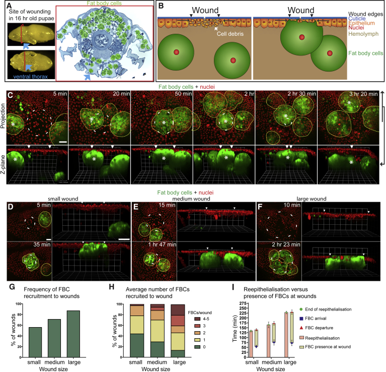
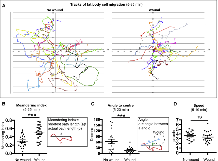
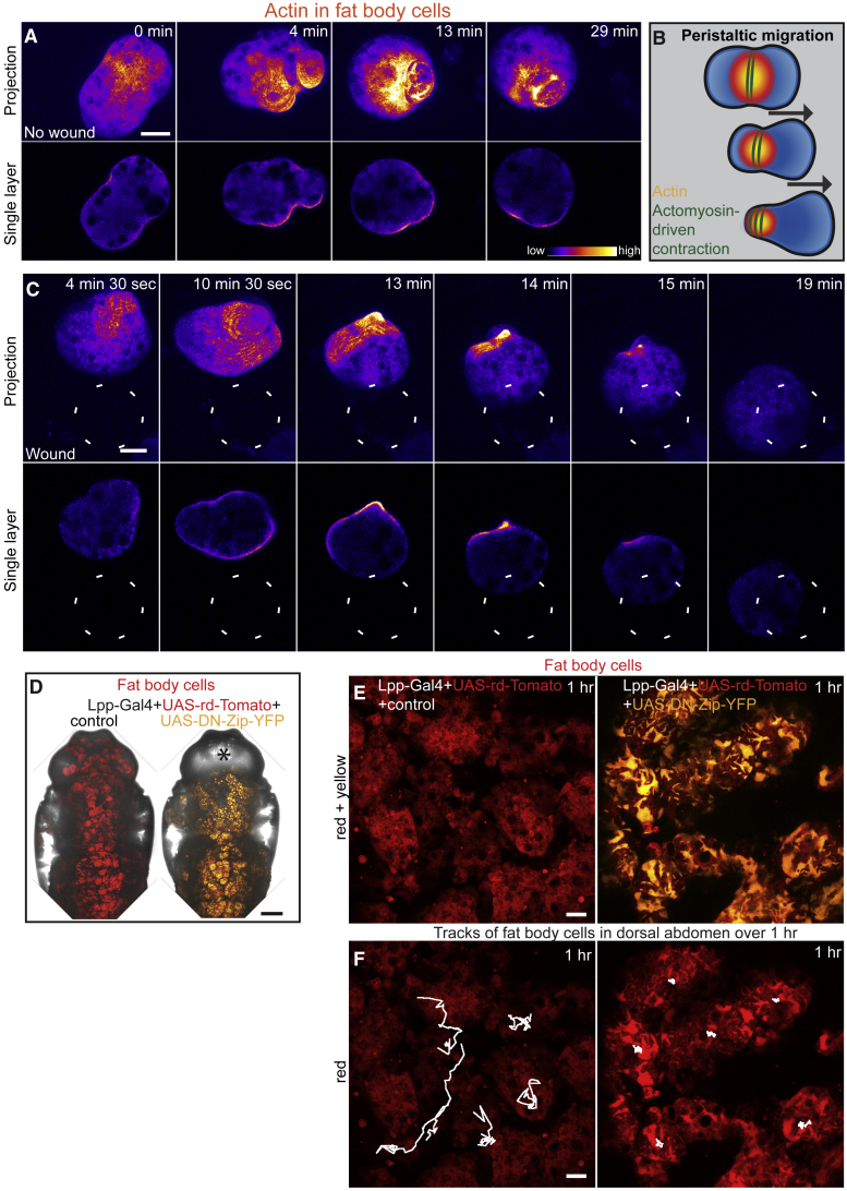
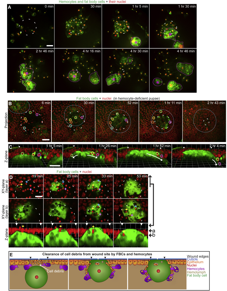
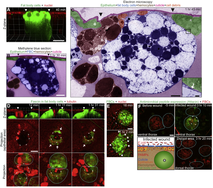
Adipocytes have many functions in various tissues beyond energy storage, including regulating metabolism, growth, and immunity. However, little is known about their role in wound healing. Here we use live imaging of fat body cells, the equivalent of vertebrate adipocytes in Drosophila, to investigate their potential behaviors and functions following skin wounding. We find that pupal fat body cells are not immotile, as previously presumed, but actively migrate to wounds using an unusual adhesion-independent, actomyosin-driven, peristaltic mode of motility. Once at the wound, fat body cells collaborate with hemocytes, Drosophila macrophages, to clear the wound of cell debris; they also tightly seal the epithelial wound gap and locally release antimicrobial peptides to fight wound infection. Thus, fat body cells are motile cells, enabling them to migrate to wounds to undertake several local functions needed to drive wound repair and prevent infections.

Keywords: Drosophila; adipocytes; antimicrobial peptides (AMPs); cell migration; fat body; hemocytes; inflammatory response; wound healing; wound infection.


Figures

Figure 1
Figure 1
FBCs Actively Migrate toward Epithelial Wounds (A) Images of pupae and methylene blue-stained section of the pupal thorax (FBCs false-colored green) showing FBC location and indicating site of laser wounding in the ventral thorax (blue arrows). (B and C) Schematic (B) and time-lapse (C) images to illustrate FBC migration to a wound (projection, C top; Z plane, B and C bottom) in a c564-Gal4+UAS-GFP+Ubq>Histone-RFP pupa (epithelial nuclei in red; FBCs in green and outlined; asterisk labels wound-associated FBCs; arrowheads indicate wound margins). See also Movie S2, first movie, and Figure S1. (D–H) Time lapse (D–F) and graphs (G and H) showing how FBCs are drawn to small, medium, and large wounds (30–60 μm, 60–90 μm, and 90–120 μm in diameter; n = 32, 12, and 15, respectively) in c564-Gal4+UAS-GFP+Ubq>Histone-RFP pupae (epithelial nuclei in red; FBCs in green and outlined). See also Movie S2, second, third, and fourth movies. (I) Graph showing duration of reepithelialization (pink bar) and FBC presence (yellow bar) in small, medium, and large wounds (n = 17, 11, and 11, respectively; genotype as in D–H). Mean ± SEM. Scale bars, 20 μm (C and D). (E) and (F) are the same magnification as (D).
Figure 2
Figure 2
FBCs Migrate to Wounds with Directional Persistence (A–D) Migration tracks of FBCs (A) and quantification of meandering index (B), angle (C), and speed (D) of FBC migration in c564-Gal4+UAS-GFP+Ubq>Histone-RFP unwounded or wounded pupae (n = 20 and 20); only analyzing cells that passed through a circular area of 25-μm radius from center within 30-min time window. See also Figure S2. Mean ± SEM. ns, p > 0.05; ∗∗∗p < 0.001 (Student's t test).
Figure 3
Figure 3
FBCs Actively Migrate toward Wounds Using a Novel Actomyosin-Driven Peristaltic Mode of Motility (A–C) Time lapse (projection at top, single plane beneath) showing actin dynamics in FBCs within unwounded (A) or wounded (C) Lpp-Gal4+UAS-GMA pupae (GMA shown in ImageJ LUT Fire). Location of the wound indicated by white dashed circle. Schematic illustrating peristaltic migration (B). See also Movie S3. (D–F) Low-magnification (D) and high-magnification (E and F) images of Lpp-Gal4+UAS-rd-Tomato+control or +UAS-DN-Zip-YFP pupae; FBCs in red, DN-Zip-YFP in yellow; asterisk marks absence of FBCs in head (D); 1-hr migration tracks of FBCs, white lines (F). See also Movie S6 and Figure S4. Control = w67. Scale bars, 20 μm (A, C, E, and F) and 200 μm (D).
Figure 4
Figure 4
FBCs and Hemocytes Together Clear the Wound of Debris (A) Time lapse of hemocyte and FBC recruitment to a wound in a srp-Gal4+c564-Gal4+UAS-GFP+UAS-Red-Stinger pupa (hemocytes are small green cells with red nuclei and yellow asterisks; FBCs are large green cells with red nuclei and outlined; purple asterisk labels wound-associated FBCs). See also Movie S7 and Figure S3. (B and C) Time-lapse sequences of wounded srp-GMA+Ubq>Histone-RFP pupae (FBC in green and outlined; epithelial nuclei in red; colored circles highlight some nuclei of necrotic epithelial cells) expressing srp-Gal4+UAS-Reaper+tubGal80ts for 16 hr before wounding at the restrictive temperature to ablate hemocytes. See also Movie S9. (D) Time lapse (two X/Y planes at top and middle and Z plane at bottom) showing phagocytic uptake of debris by an FBC in a wounded c564-Gal4+UAS-GFP+Ubq>Histone-RFP pupa (FBCs in green; epithelial nuclei in red; colored arrows highlight some nuclei of necrotic epithelial cells to aid tracking). (E) Schematic illustrating collaborative clearance of cell debris from wound site by FBCs and hemocytes. Scale bars, 20 μm (A–C) and 10 μm (D).
Figure 5
Figure 5
FBCs Seal the Wound and Locally Produce AMPs (A) Time lapse (single frame) of wound-plugging by an FBC in a c564-Gal4+UAS-GFP+Ubq>Histone-RFP pupa (FBC in green; epithelial nuclei in red). (B and C) Methylene blue-stained resin section (B) and transmission electron microscopy (C) images of FBC plugging the wound (different cell types are shown false-colored, as indicated). (D) Time lapse of lamellipodia formation by FBCs at the wound of a c564-Gal4+UAS-GFP-Fascin+Ubq>RFP-tubulin pupa (epithelium in red; Fascin in green, Fascin-rich protrusions indicated with arrows). See also Movie S5. (E) Time lapse of FBC blebbing at a wound in a c564-Gal4+UAS-GFP+Ubq>Histone-RFP pupa (epithelial nuclei in red; FBCs in green; blebs indicated with arrows). See also Movie S2, second movie. (F) Time lapse of local Attacin expression in FBCs in an Attacin>GFP+Lpp-Gal4+UAS-myr-td-Tom pupa after wounding and exposure to RFP-E. coli for 5 min (FBCs in red and outlined; Attacin expression, green). Schematic illustrating experimental setup. See also Movie S10. Scale bars, 20 μm (A, B, and D–F), 5 μm (C) and 500 nm (C, insert).

Comment in

References

    1. Antunes M., Pereira T., Cordeiro J.V., Almeida L., Jacinto A. Coordinated waves of actomyosin flow and apical cell constriction immediately after wounding. J. Cell Biol. 2013;202:365–379. - PMC - PubMed
    1. Barolo S., Castro B., Posakony J.W. New Drosophila transgenic reporters: insulated P-element vectors expressing fast-maturing RFP. Biotechniques. 2004;36:436–440. 442. - PubMed
    1. Barry N.P., Bretscher M.S. Dictyostelium amoebae and neutrophils can swim. Proc. Natl. Acad. Sci. USA. 2010;107:11376–11380. - PMC - PubMed
    1. Basto R., Brunk K., Vinadogrova T., Peel N., Franz A., Khodjakov A., Raff J.W. Centrosome amplification can initiate tumorigenesis in flies. Cell. 2008;133:1032–1042. - PMC - PubMed
    1. Beller M., Bulankina A.V., Hsiao H.-H.H., Urlaub H., Jäckle H., Kühnlein R.P. PERILIPIN-dependent control of lipid droplet structure and fat storage in Drosophila. Cell Metab. 2010;12:521–532. - PubMed

Publication types

MeSH terms