. 2018 Oct;68(4):1574-1588.
doi: 10.1002/hep.29857. Epub 2018 May 21.

Intestine farnesoid X receptor agonist and the gut microbiota activate G-protein bile acid receptor-1 signaling to improve metabolism

Affiliations

Intestine farnesoid X receptor agonist and the gut microbiota activate G-protein bile acid receptor-1 signaling to improve metabolism

Preeti Pathak et al. Hepatology. 2018 Oct.

Abstract

Bile acids activate farnesoid X receptor (FXR) and G protein-coupled bile acid receptor-1 (aka Takeda G protein-coupled receptor-5 [TGR5]) to regulate bile acid metabolism and glucose and insulin sensitivity. FXR and TGR5 are coexpressed in the enteroendocrine L cells, but their roles in integrated regulation of metabolism are not completely understood. We reported recently that activation of FXR induces TGR5 to stimulate glucagon-like peptide-1 (GLP-1) secretion to improve insulin sensitivity and hepatic metabolism. In this study, we used the intestine-restricted FXR agonist fexaramine (FEX) to study the effect of activation of intestinal FXR on the gut microbiome, bile acid metabolism, and FXR and TGR5 signaling. The current study revealed that FEX markedly increased taurolithocholic acid, increased secretion of fibroblast growth factors 15 and 21 and GLP-1, improved insulin and glucose tolerance, and promoted white adipose tissue browning in mice. Analysis of 16S ribosomal RNA sequences of the gut microbiome identified the FEX-induced and lithocholic acid-producing bacteria Acetatifactor and Bacteroides. Antibiotic treatment completely reversed the FEX-induced metabolic phenotypes and inhibited taurolithocholic acid synthesis, adipose tissue browning, and liver bile acid synthesis gene expression but further increased intestinal FXR target gene expression. FEX treatment effectively improved lipid profiles, increased GLP-1 secretion, improved glucose and insulin tolerance, and promoted adipose tissue browning, while antibiotic treatment reversed the beneficial metabolic effects of FEX in obese and diabetic mice.

Conclusion: This study uncovered a mechanism in which activation of intestinal FXR shaped the gut microbiota to activate TGR5/GLP-1 signaling to improve hepatic glucose and insulin sensitivity and increase adipose tissue browning; the gut microbiota plays a critical role in bile acid metabolism and signaling to regulate metabolic homeostasis in health and disease. (Hepatology 2018).


Conflict of interest statement

Disclosures: No potential conflicts of interest relevant to this article were reported.

Figures

Fig. 1
Fig. 1
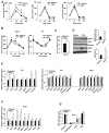
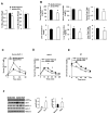
The intestine-restricted FXR agonist FEX stimulated GLP-1 secretion, hepatic insulin sensitivity and adipose tissue browning in mice. Wild-type C57BL/6J mice were gavaged with FEX (50 mg/Kg, n=10), or vehicle (0.2% DMSO in PBS, n=10) for 7 days. For oral glucose tolerance testing, the mice were fasted for 6 h and gavaged with glucose (2 g/Kg). (A) Effect of FEX on GLP-1 secretion in wild-type, Fxr−/− and Tgr5−/− mice. (B) Oral glucose tolerance test (left) and insulin tolerance test (right) of wild-type mice treated with FEX. (C) ELISA assay of serum FGF21 levels in FEX-treated wild-type mice. (D) Effect of FEX treatment on phosphorylation of liver AKT and acetyl-CoA carboxylase. Total and phosphorylated AKT and ACC were assayed by immunoblot analysis of liver extracts of vehicle-treated (n=3) and FEX-treated (n=4) mice. (E) Real-time PCR analysis of the effect of FEX treatment on mRNA expression of browning factors in inguinal white adipose tissue (iWAT) of wild-type, Fxr−/− and Tgr5−/− mice. (F) Real-time PCR analysis of the effect of FEX treatment on mRNA expression of FXR targets in mouse ileum. (G) Effect of FEX treatment on bile acid contents in intestine, gallbladder and liver and total bile acid pool size in wild-type mice. Results were expressed as mean ± standard error. An “*” indicates statistically significant difference between treated vs. vehicle control, p ≤ 0.05. Student’s t-test was used for statistical analysis.
Fig. 2
Fig. 2
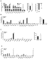
FEX treatment altered bile acid composition in wild-type mice. Wild-type C57BL/6J mice were orally gavaged with the FXR agonist FEX (50 mg/Kg, n=8), or vehicle (0.2% DMSO in PBS, n=7) as indicated for 9 days. Mice were fasted for 6 h and killed. (A) FEX altered bile acid concentration in gallbladder bile. Bile acids were extracted from 2 μl gallbladder bile from FEX-treated and vehicle control mice. Hydrophobicity index of gallbladder bile was calculated by multiplying the hydrophobicity index of individual bile acids by the concentration (mM) of the individual bile acids in the gallbladder. (B) Ileum bile acid composition. (C) Colon bile acid composition. (D) Serum bile acid composition. Hydrophobicity index used: TCA= 0, T7α-MCA= −0.84, Tβ-MCA= −0.78, THDCA= −0.37, Tγ-MCA and Tω-MCA = −0.33, TUDCA= −0.27, TCDCA= 0.46, TDCA= 0.59, TLCA= 1. Results were expressed as mean ± standard error. Results were expressed as mean ± standard error. An “*” indicates statistically significant difference (p ≤ 0.05) treated vs. vehicle control. An “**” indicates statistically significant difference between treated vs. vehicle control, p ≤ 0.01. Student’s t-test was used for statistical analysis.
Fig. 3
Fig. 3
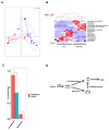
Gut microbiota analysis. Gut microbes were extracted according the described methods after FEX treatment. FEX significantly altered the composition of the gut microbiota. (A) Generalized unifrac analysis of the whole gut microbiota population between vehicle and FEX-treated mice. Generalized unifrac combines weighted and unweighted unifrac analysis to incorporate both abundant and rare taxonomies, respectively. Significance was obtained via the R package Adonis, which is a permutational multivariate analysis of variance that is designed for distance matrices. (B) Effect of FEX on the genera of the gut microbiome. Z-scores are used to illustrate significantly different genera (p ≤ 0.05) between vehicle and FEX-treated mice. Samples were allowed to cluster under the R package heatmap.2’s hclust function. The dendrogram shows related samples. (C) Two genera, Acetatifactor and Bacteroides, were shown to increase after FEX treatment. (D) Gut bacteria induce LCA production to stimulate TGR5 signaling. Bile salt hydrolase de-conjugates TCDCA to CDCA, which is converted to LCA by bacterial 7α-dehydroxylase. CDCA can be epimerized to UDCA, which is converted to LCA by bacterial 7β-dehydroxylase. LCA activates TGR5 in the intestine. An “*” indicates statistically significant difference (p ≤ 0.05) between treated vs. vehicle control.
Fig. 4
Fig. 4
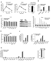
Antibiotics prevented FEX-induced metabolic phenotypes in wild-type mice. Wild-type C57BL/6J mice were given a mixture of antibiotics (ABX) in drinking water, containing ampicillin (1 g/L), vancomycin (500 mg/L), neomycin sulfate (g/L), and metronidazole (1 g/L) for 30 days. In the last week of antibiotic treatment, mice were orally gavaged with FEX (50 mg/Kg, n=10 each group), or vehicle (0.2% DMSO in PBS, n=10 each group) for 7 days. (A) FEX did not affect GLP-1 secretion in ABX-treated mice (left). Serum GLP-1 levels were assayed over 60 min in FEX + antibiotic (FEX+ABX) and vehicle + antibiotic (Vehicle+ABX) treated-mice. FEX did not stimulate oral glucose tolerance in ABX-treated mice (middle). FEX did not increase serum FGF21 levels in ABX-treated mice (right). (B) FEX reduced gallbladder and liver bile acid content, resulting in a significantly reduced bile acid pool.. (C) FEX treatment reduced FXR target gene mRNA expression (left panel) and CYP7A1 and CYP8B1 protein expression (right panel) in the liver of ABX-treated mice. (D) Effect of FEX treatment on mRNA expression levels of FXR targets in the ileum of mice treated with ABX. (E) Effect of FEX treatment on mRNA expression levels of browning factors in eWAT of ABX-treated mice. (F). FEX did not alter bile acid composition in gallbladder bile of ABX-treated mice. (G). FEX did not alter bile acid composition in serum of ABX-treated mice. Results in all panels were expressed as mean ± standard error. An “*” indicates statistically significant difference determined by Student’s t-test (p ≤ 0.05), FEX+ABX-treated vs. vehicle + ABX-treated wild-type mice.
Fig. 5
Fig. 5
FEX increases GLP-1 secretion and improves insulin sensitivity in db/db mice. db/db mice (n=6 in each group) were treated by oral gavage with FEX (30 mg/Kg) + sitagliptin (3 mg/Kg) or vehicle (0.2% DMSO) + sitagliptin (3 mg/Kg) for 9 days. (A) Effect of FEX treatment on fat mass and lean mass in db/db mice. (B) Effect of FEX treatment on serum lipid profiles in db/db mice. (C) GLP-1 secretion assay of FEX in db/db mice. (D) Oral glucose tolerance assay of effect of FEX in db/db mice. (E) Insulin tolerance test of effect of FEX in db/db mice. (F) Effect of FEX on phosphorylation of liver AKT473 and ACC-1 in db/db mice. Each lane represents one mouse (n=3 per group). An “*” indicates statistically significant difference between treated vs. vehicle control (p ≤ 0.05), determined by Student’s t-test.
Fig. 6
Fig. 6
Effect of FEX in liver and adipose tissue gene mRNA expression in db/db mice (A) Effect of FEX on intestinal FXR target gene mRNA expression (left), hepatic gluconeogenic gene mRNA expression, serum FGF21. (B) Effect of FEX on brown adipose activation genes in brown adipose tissue (BAT). (C) Effect of FEX on adipose browning factors in subcutaneous WAT (sWAT), epididymal WAT (eWAT), and inguinal WAT (iWAT). (D) UCP-1 protein expression in the inguinal fat. An “*” indicates statistically significant difference between treated vs. vehicle control (p ≤ 0.05), determined by Student’s t-test.

Comment in

  • Bile Acids, Microbiota, and Metabolism.
    Taylor SA, Green RM. Taylor SA, et al. Hepatology. 2018 Oct;68(4):1229-1231. doi: 10.1002/hep.30078. Hepatology. 2018. PMID: 29729182 Free PMC article. No abstract available.

Similar articles

Cited by

References

    1. Lefebvre P, Cariou B, Lien F, Kuipers F, Staels B. Role of bile acids and bile Acid receptors in metabolic regulation. Physiol Rev. 2009;89:147–191. - PubMed
    1. Li T, Chiang JY. Bile acid signaling in metabolic disease and drug therapy. Pharmacol Rev. 2014;66:948–983. - PMC - PubMed
    1. Chiang JY. Bile acids: regulation of synthesis. J Lipid Res. 2009;50:1955–1966. - PMC - PubMed
    1. Inagaki T, Choi M, Moschetta A, Peng L, Cummins CL, McDonald JG, Luo G, et al. Fibroblast growth factor 15 functions as an enterohepatic signal to regulate bile acid homeostasis. Cell Metab. 2005;2:217–225. - PubMed
    1. Jiang C, Xie C, Li F, Zhang L, Nichols RG, Krausz KW, Cai J, et al. Intestinal farnesoid X receptor signaling promotes nonalcoholic fatty liver disease. J Clin Invest. 2015;125:386–402. - PMC - PubMed

Publication types

MeSH terms

Substances