

Review
doi: 10.1104/pp.17.01776.
Epub 2018 Feb 27.
Monitoring Polysaccharide Dynamics in the Plant Cell Wall
Affiliations
- PMID: 29487120
- PMCID: PMC5884611
- DOI: 10.1104/pp.17.01776
Item in Clipboard
Review
Monitoring Polysaccharide Dynamics in the Plant Cell Wall
Plant Physiol.
2018 Apr.
Abstract
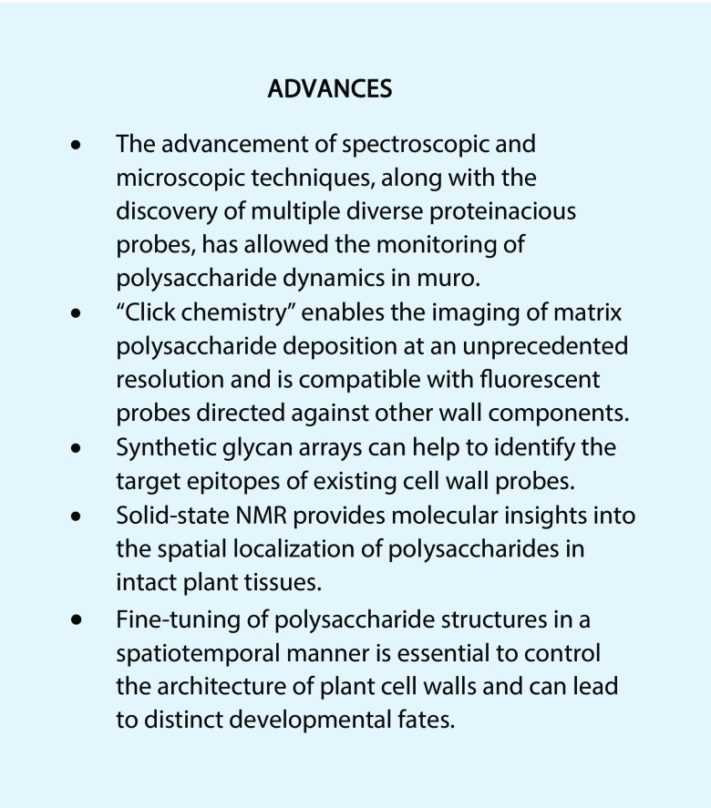
New technologies reveal the deposition and remodeling of plant cell wall polysaccharides and their impact on plant development.
Figures

Major classes of probes and key steps to image cell wall polysaccharides. A, Illustration of the steps required prior to microscopy (represented by the magnifying glasses) for different types of probes. Secondary antibodies (2° mAb) are commercially available and should be selected based on the final application (electron versus light microscopy) and the available equipment (e.g. fluorescence filters). Probes in colored boxes are exemplified in B to G. B and C, Sites of wall polysaccharide synthesis in Arabidopsis cotyledons expressing the yellow FP markers wave_22Y and wave_138Y, respectively (Geldner et al., 2009). FP signal and chloroplast intrinsic fluorescence are shown using Orange Hot and Cyan Hot look-up tables in Fiji (Schindelin et al., 2012). D to G, Mucilage architecture of an S4B-stained and mAb-labeled (CCRC-M36 and Alexa Fluor 488 2° mAb) Arabidopsis seed. D shows a segment of a whole seed (black) that has been brought into contact with water. The only clearly visible structures are columellae (C). S4B staining reveals cellulosic rays in E, and the antibody CCRC-M36 shows the deposition of rhamnogalacturonan in F. The comparison of the overlay G with D exemplifies the usefulness of dyes and labels. Bars = 25 µm (B–G).
References
-
- Atmodjo MA, Hao Z, Mohnen D (2013) Evolving views of pectin biosynthesis. Annu Rev Plant Biol 64: 747–779 - PubMed
-
- Barnes WJ, Anderson CT (2018) Release, recycle, rebuild: cell-wall remodeling, autodegradation, and sugar salvage for new wall biosynthesis during plant development. Mol Plant 11: 31–46 - PubMed
Publication types
MeSH terms
Substances
LinkOut - more resources
Full Text Sources
Other Literature Sources
