. 2018 Apr 20;293(16):6052-6063.
doi: 10.1074/jbc.RA117.000731. Epub 2018 Feb 27.

Mass spectrometric evidence for neuropeptide-amidating enzymes in Caenorhabditis elegans

Affiliations

Mass spectrometric evidence for neuropeptide-amidating enzymes in Caenorhabditis elegans

Sven Van Bael et al. J Biol Chem. .

Abstract

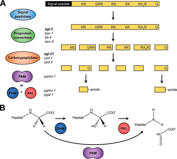
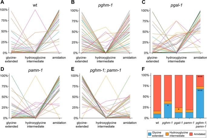
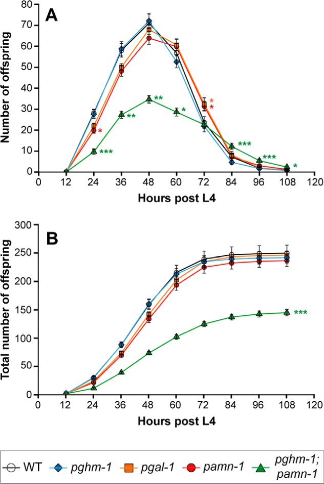
Neuropeptides constitute a vast and functionally diverse family of neurochemical signaling molecules and are widely involved in the regulation of various physiological processes. The nematode Caenorhabditis elegans is well-suited for the study of neuropeptide biochemistry and function, as neuropeptide biosynthesis enzymes are not essential for C. elegans viability. This permits the study of neuropeptide biosynthesis in mutants lacking certain neuropeptide-processing enzymes. Mass spectrometry has been used to study the effects of proprotein convertase and carboxypeptidase mutations on proteolytic processing of neuropeptide precursors and on the peptidome in C. elegans However, the enzymes required for the last step in the production of many bioactive peptides, the carboxyl-terminal amidation reaction, have not been characterized in this manner. Here, we describe three genes that encode homologs of neuropeptide amidation enzymes in C. elegans and used tandem LC-MS to compare neuropeptides in WT animals with those in newly generated mutants for these putative amidation enzymes. We report that mutants lacking both a functional peptidylglycine α-hydroxylating monooxygenase and a peptidylglycine α-amidating monooxygenase had a severely altered neuropeptide profile and also a decreased number of offspring. Interestingly, single mutants of the amidation enzymes still expressed some fully processed amidated neuropeptides, indicating the existence of a redundant amidation mechanism in C. elegans All MS data are available via ProteomeXchange with the identifier PXD008942. In summary, the key steps in neuropeptide processing in C. elegans seem to be executed by redundant enzymes, and loss of these enzymes severely affects brood size, supporting the need of amidated peptides for C. elegans reproduction.

Keywords: Caenorhabditis elegans (C. elegans); PAL; PAM; PHM; amidation; copper monooxygenase; mass spectrometry (MS); neuropeptide; peptides; peptidomics.


Conflict of interest statement

The authors declare that they have no conflicts of interest with the contents of this article

Figures

Figure 1.
Figure 1.
Neuropeptide-processing pathway. Neuropeptides are synthesized as large preproproteins that require post-translational processing, and as exemplified in A, the signal peptide is cleaved upon entry into the secretory pathway by a signal peptidase. Subsequently, a proprotein convertase (mainly egl-3, but also kpc-1, bli-4, and aex-5 in C. elegans) cleaves the remaining part of the precursor protein at specific motifs containing basic amino acids (KR, RR, RK, KK, or RXnR with n = 2, 4, 6, or 8). These residues are then removed by a carboxypeptidase (EGL-21, CPD-1 and CPD-2 in C. elegans) to yield the cleaved peptide. Finally, the carboxyl-terminal glycine residue, if present, is transformed into an amide. B, carboxyl-terminal amidation involves two steps: hydroxylation of the glycine α-carbon by a PHM, followed by a cleavage reaction performed by a PAL. This will generate a glyoxylate molecule and the α-amidated peptide. In vertebrates, these two enzymatic activities are contained in one bifunctional enzyme, PAM.
Figure 2.
Figure 2.
Inter-phyla sequence comparison of bifunctional PAM and monofunctional PHM enzymes. Multiple sequence alignments of the PHM domain of PAM enzymes are from H. sapiens (NP_000910.2), the mouse M. musculus (P97467.2), the zebrafish D. rerio (XP_699436.4), the African clawed frog X. laevis (NP_001079520.2), the marine snail A. californica (AAF67216.1), the sea anemone C. parasitica (Q9GQN2), and C. elegans (P83388.2) with PHM enzymes from C. elegans (Q95XM2.1), the parasitic platyhelminth S. mansoni (AAO18222.1), the planarian D. japonica (BAD98846.1), the fruit fly D. melanogaster (AAF47127.1), and C. parasitica (AAG24505.1). Whereas eight canonical cysteine residues (numbered 1–8, residues marked in red) that form four putative disulfide bridges are present in PHM enzymes and in the PHM domains of the bifunctional PAM enzymes, cysteines 1 and 3 are missing in C. elegans PAM. All residues marked with an asterisk are involved in the catalytic function of PHM and are highly conserved throughout the species. One copper ion is bound by three histidine residues, marked in green. The second catalytic copper ion is held in place by two histidines and one methionine (marked in blue). The arginine, asparagine, and tyrosine residues marked in orange are part of the substrate-binding site and interact with the peptide to be amidated (14).
Figure 3.
Figure 3.
Inter-phyla sequence comparison of bifunctional PAM and monofunctional PAL enzymes. Multiple sequence alignments of the PAL domain of PAM enzymes are from H. sapiens (NP_000910.2), the mouse M. musculus (P97467.2), the zebrafish D. rerio (XP_699436.4), the African clawed frog X. laevis (NP_001079520.2), the marine snail A. californica (AAF67216.1), the sea anemone C. parasitica (Q9GQN2), and C. elegans (P83388.2) with PAL enzymes from C. elegans (CCD69816.1), the parasitic platyhelminth S. mansoni (ACN42951.1), and the fruit fly D. melanogaster (ACJ13179.1 and AAF47043.2). Two conserved cysteines that form a disulfide bridge are numbered and marked in red. Critical residues for catalytic activity are marked with an asterisk or colon, denoting perfectly conserved or partially conserved residues, respectively. The residues involved in the binding of a catalytic zinc ion are marked in green and are highly conserved throughout species. Residues marked in yellow are responsible for coordination of a calcium ion. These seem to lack a high degree of conservation in other species. Although one Ca2+-binding leucine residue is still present in C. elegans PAL, they appear to be completely absent in C. elegans PAM. Key active site residues are marked in orange. Both the catalytic arginine and tyrosine are highly conserved, and the methionine residue that binds and positions the substrate shows a much lower conservation. The low conservation of this methionine and the Ca2+-binding residues in different species were also observed by Atkinson et al. (18).
Figure 4.
Figure 4.
Modification states of separate identified neuropeptides (A–E) and distribution of neuropeptide modification states (F) in WT and mutant animals. Each individual line corresponds to an identified neuropeptide (52 in total). The y axis represents the mean values of normalized relative peptide abundances. The majority of neuropeptides in wildtype (wt) (A) are detected as carboxyl-terminal amidated peptides. Mutating pghm-1 seems to disrupt normal amidation, because more neuropeptides are detected with the carboxyl-terminal glycine still present (B). Knockout of pgal-1 seems to have only a small effect on the amount of amidated neuropeptides (C); however, an increase in hydroxyglycine intermediates can be seen. Inactivation of pamn-1 has no severe effects on amidation (D). Finally, mutating both pghm-1 and pamn-1 displays the most severe amidation effects (E). The majority of neuropeptides are found with their carboxyl-terminal glycine still present. When looking at the modification state distribution for all 52 peptides (F), WT animals indeed show a high occurrence of amidation (84.4%) and only low amounts of glycine-extended peptides. Although knockout of pghm-1 results in a rise of glycine-extended peptides (*, p < 0.05), other single mutants seem to be less affected. Although pgal-1 shows a significant increase in hydroxyglycine intermediates (*, p < 0.05), it still contains a high amount of amidated peptides. pamn-1 knockout does not seem to have any major effects on peptide amidation, because it closely resembles WT. A clear effect is seen in the pghm-1;pamn-1 double mutant, where the occurrence of amidated peptides collapses (***, p < 0.001) and is concomitant with a rise in glycine-extended peptides (***, p < 0.001). Because the effect in the double mutant is more severe than the additive effects of each single mutant, this may suggest that pghm-1 and pamn-1 are interchangeable to a certain degree. Data are derived from 52 identified peptides, present in all mutants. For each mutant, three (WT, pgal-1, and pamn-1) or two (pghm-1 and pghm-1;pamn-1) replicates were used. For more detail, including data of individual replicates and annotation of all individual neuropeptides, see Figs. S2 and S3.
Figure 5.
Figure 5.
Egg-laying profile (A) and total brood size (B) of WT (n = 27), pamn-1 (n = 35), pgal-1 (n = 32), pghm-1 (n = 22), and pghm-1;pamn-1 (n = 21) mutant worms. The single mutants do not show any severe defects in egg-laying behavior nor a difference in total brood size. Inactivation of both pghm-1 and pamn-1, however, results in a severely diminished brood size that is reduced by 42% compared with WT (*, p < 0.05; **, p < 0.01; ***, p < 0.001).
Figure 6.
Figure 6.
Modification states of separate identified neuropeptides (A and B) and distribution of neuropeptide modification states (C) in WT and tbh-1 animals. Each individual line corresponds to an identified neuropeptide (36 in total). The y axis represents the mean values of normalized relative peptide abundances. The majority of all neuropeptides are detected as carboxyl-terminally amidated peptides in both WT (A) as well as in tbh-1 mutants (B). This is also apparent in the overall modification state distribution of all 36 peptides (C), where no significant differences were observed between WT and tbh-1 mutants. For more detail, including data of individual replicates and annotation of all individual neuropeptides, see Fig. S6.
Figure 7.
Figure 7.
Co-localization of pghm-1, pgal-1, and pamn-1. Expression of pghm-1::GFP, pgal-1::CFP, and pamn-1::mCherry fusion constructs was monitored in young adults by confocal imaging in multitrack mode. Co-localization of the three reporter constructs could be observed in most of the cell bodies of the nerve ring in the head (upper panels) and the tail ganglia (lower panels).

Similar articles

Cited by

References

    1. Li C., and Kim K. (2010) Neuropeptide gene families in Caenorhabditis elegans. Adv. Exp. Med. Biol. 692, 98–137 10.1007/978-1-4419-6902-6_6 - DOI - PubMed
    1. Husson S. J., Reumer A., Temmerman L., De Haes W., Schoofs L., Mertens I., and Baggerman G. (2014) Worm peptidomics. EuPA Open Proteomics 3, 280–290 10.1016/j.euprot.2014.04.005 - DOI
    1. Hobert O. (2013) The neuronal genome of Caenorhabditis elegans. WormBook 2013 10.1895/wormbook.1.161.1 - DOI - PMC - PubMed
    1. Husson S. J., Clynen E., Baggerman G., Janssen T., and Schoofs L. (2006) Defective processing of neuropeptide precursors in Caenorhabditis elegans lacking proprotein convertase 2 (KPC-2/EGL-3): mutant analysis by mass spectrometry. J. Neurochem. 98, 1999–2012 10.1111/j.1471-4159.2006.04014.x - DOI - PubMed
    1. Husson S. J., Janssen T., Baggerman G., Bogert B., Kahn-Kirby A. H., Ashrafi K., and Schoofs L. (2007) Impaired processing of FLP and NLP peptides in carboxypeptidase E (EGL-21)-deficient Caenorhabditis elegans as analyzed by mass spectrometry. J. Neurochem. 102, 246–260 10.1111/j.1471-4159.2007.04474.x - DOI - PubMed

Publication types

MeSH terms