. 2018 Mar 13;115(11):E2538-E2545.
doi: 10.1073/pnas.1722050115. Epub 2018 Feb 27.

Structural basis of transcriptional stalling and bypass of abasic DNA lesion by RNA polymerase II

Affiliations

Structural basis of transcriptional stalling and bypass of abasic DNA lesion by RNA polymerase II

Wei Wang et al. Proc Natl Acad Sci U S A. .

Abstract

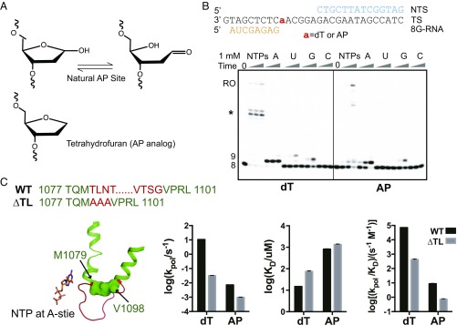
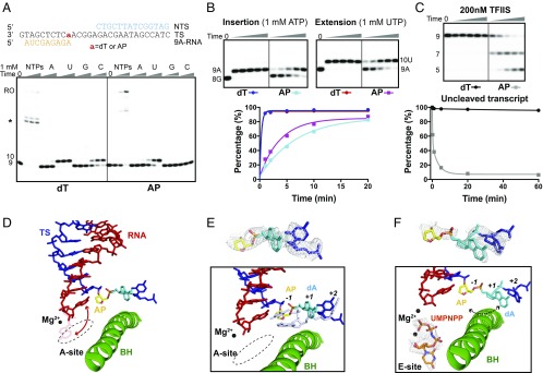
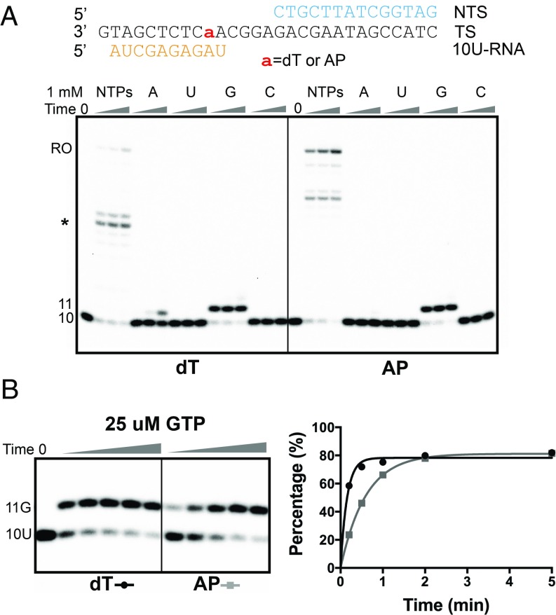
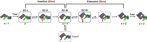
Abasic sites are among the most abundant DNA lesions and interfere with DNA replication and transcription, but the mechanism of their action on transcription remains unknown. Here we applied a combined structural and biochemical approach for a comprehensive investigation of how RNA polymerase II (Pol II) processes an abasic site, leading to slow bypass of lesion. Encounter of Pol II with an abasic site involves two consecutive slow steps: insertion of adenine opposite a noninstructive abasic site (the A-rule), followed by extension of the 3'-rAMP with the next cognate nucleotide. Further studies provided structural insights into the A-rule: ATP is slowly incorporated into RNA in the absence of template guidance. Our structure revealed that ATP is bound to the Pol II active site, whereas the abasic site is located at an intermediate state above the Bridge Helix, a conserved structural motif that is cirtical for Pol II activity. The next extension step occurs in a template-dependent manner where a cognate substrate is incorporated, despite at a much slower rate compared with nondamaged template. During the extension step, neither the cognate substrate nor the template base is located at the canonical position, providing a structural explanation as to why this step is as slow as the insertion step. Taken together, our studies provide a comprehensive understanding of Pol II stalling and bypass of the abasic site in the DNA template.

Keywords: RNA polymerase II; abasic sites in DNA; transcription pausing; transcriptional mutagenesis; translesion transcription.


Conflict of interest statement

The authors declare no conflict of interest.

Figures

Fig. 1.
Fig. 1.
Strong transcription pausing opposite the AP site at the insertion step. (A) Natural AP site is in equilibrium between the closed sugar ring hemiacetal and the open aldehyde forms. The synthetic AP analog tetrahydrofuran used in this study is shown. (B) Gel analysis showed Pol II transcription elongation was stalled across an AP site. The concentration of NTP is 1 mM. Time points are 1 min, 5 min, and 30 min. The bands marked as * are sequence-specific pausings. (C) Comparison of catalytic rate constants (kpol), substrate dissociation constants (KD), and specificity constants (kpol/KD) of ATP incorporation opposite the AP template by wild-type (WT) and ΔTL Pol II, respectively. Left shows the amino sequences (central portion) and ΔTL of Pol II. The central portion of the TL motif (T1080-G1097) that is involved in conformational changes and NTP substrate interaction is deleted and replaced by three alanines in ΔTL.
Fig. 2.
Fig. 2.
Structural analysis of transcription stalling against the AP site. (A) Crystal structure of Pol II EC with an AP site at the i + 1 position (EC-I). Upper shows the AP site is kept as an intermediate state by Rpb1 R337. Lower shows the comparison of the AP site with the undamaged dT template (silver, PDB ID code 4BY1). (B) Overall structure of Pol II EC-II containing an AP site template and AMPCPP. Pol II is shown from the side view as a ribbon model in gray, with the Bridge Helix (BH) highlighted in green and Rpb2 omitted for clarity. The nucleic acids are shown as stick models: template DNA, TS, blue; RNA, red. The AP site and AMPCPP are highlighted as yellow and orange, respectively. The colors are consistent in all structural figures unless specified otherwise. Lower shows 2Fo − Fc maps (gray) of the AP site and AMPCPP are contoured at 1.0 σ. (C) Upper shows the detailed interaction between AMPCPP and Pol II. Mg2+ and H2O are shown as black and blue dots, respectively. Lower shows the comparison of the AP:AMPCPP pair with the canonical dT:AMPCPP pair (silver, PDB ID code 4BY1).
Fig. 3.
Fig. 3.
The AP site induced strong transcription pausing at the extension step. (A) Transcription assay revealed that the AP site causes transcriptional stalling in the extension step. Although the RNA extension after the AP site is template-dependent, it occurs with a substantially slower rate than the extension on the nondamaged template. The concentration of NTP is 1 mM. Time points are 1 min, 5 min, and 30 min. (B) Comparison of nucleotide incorporation efficiency at the insertion and extension steps for the AP-containing template. The concentration of NTP is 1 mM. Time points are 1 min, 2 min, 5 min, 10 min, and 20 min. For AP-containing Pol II EC, the kobs of ATP incorporation at the insertion step is 0.15 ± 0.01 min−1, and the kobs of UTP incorporation at the extension step is 0.27 ± 0.02 min−1. (C) The presence of the AP site at the DNA template promotes further backtracking (7-mer product), revealed by TFIIS-stimulated transcript cleavage assay. The 5-mer RNA product is caused by an unstable, shorter RNA/DNA hybrid (n < 8). Time points are 15 s, 1 min, 5 min, 20 min, and 60 min. The concentration of TFIIS is 200 nM. Quantitative analysis shows that kobs is 4.5 ± 0.4 min−1 for the AP-lesion template. (D) Crystal structure of Pol II EC with an AP site at the i − 1 position (EC-III). The 3′ RNA nucleotide (9A) is located at the canonical i − 1 position “pairing” with the AP site. One potential frayed conformation of 9A is shown as a red stick with 80% transparency. Red arrow represents a possible switch between two conformations. The 5′ nucleotide (dA) next to the AP site is colored cyan. (E) The comparison of the AP site with the undamaged dT template (silver, PDB ID code 2NVZ). A lack of base-stacking effect of an AP site causes difficulty of the 5′ nucleotide (dA) locating at the canonical i + 1 position. (F) Crystal structure of Pol II EC with an AP site at the i − 1 position and soaking UMPNPP (EC-IV). UMPNPP is located at the E-site without pairing with dA at the i + 1 position (dashed arrow represents its potential positonal changes). The 2Fo − Fc maps (gray) of the AP site and UMPNPP are contoured at 1.0 σ.
Fig. 4.
Fig. 4.
Pol II can elongate beyond the AP site after two consecutive stallings at the insertion and extension steps. (A) Transcription assay shows that an upstream AP site has no significant effect on transcription elongation after the extension step under 1 mM NTP concentration. Time points are 1 min, 5 min, and 30 min. (B) Under lower GTP concentration (25 μM), the upstream AP site only causes weak transcription pausing after the extension step. Time points are 12 s, 30 s, 1 min, 2 min, and 5 min. For the undamged template, kobs,dT = 6.5 ± 0.5 min−1; for the AP-containing template, kobs,AP = 1.68 ± 0.04 min−1.
Fig. 5.
Fig. 5.
Model of AP lesion-induced stalling and bypass by Pol II based on current biochemical and structural data. The RNA and template DNA are highlighted in red and dark blue, respectively. The AP site and pairing ATP (or AMP) are colored as yellow and orange, respectively. The Bridge Helix (BH) is shown in green. Models in solid box represent the solved crystal structures (EC I–IV). The dashed boxes show inferred models based on biochemical data. Briefly, Pol II is not stalled until it reaches the AP site. Two consective bond formation steps (insertion and extension) are strongly inhibited by the AP site. The structures of EC-I (without NTP) and EC-II (with bound NTP) depict the states where Pol II encounters the AP site (register n). In the first stalled state (insertion step), while the RNA–DNA hybrid is in a canonical posttranslocation register, the AP site occupies an intermediate state above the Bridge Helix and is not loaded to the active site (EC-I). ATP binds opposite the AP site (A-rule) in EC-II with the TL remaining in an open conformation, which is consistent with the slow rate of nucleotide addition in EC-II (EC-II → EC-III step). The second stalled state (EC-III) is characterized by a significant structural flexibility for both 3′ AMP and the 5′ template nucleotide adjacent to the AP site. This is due to a lack of stacking interactions and base pair restraint, which interferes with binding of the incoming cognate UTP. As a result, UTP largely occupies the flipped-out conformation in the E-site (EC-IV). Once the slow extension step is completed, Pol II rapidly escapes from the lesion, with the rate similar to transcription on the nondamaged template (n + 2 to n + 3).

Similar articles

Cited by

References

    1. Lindahl T. Instability and decay of the primary structure of DNA. Nature. 1993;362:709–715. - PubMed
    1. Nakamura J, et al. Highly sensitive apurinic/apyrimidinic site assay can detect spontaneous and chemically induced depurination under physiological conditions. Cancer Res. 1998;58:222–225. - PubMed
    1. Loeb LA, Preston BD. Mutagenesis by apurinic/apyrimidinic sites. Annu Rev Genet. 1986;20:201–230. - PubMed
    1. Goodman MF. Error-prone repair DNA polymerases in prokaryotes and eukaryotes. Annu Rev Biochem. 2002;71:17–50. - PubMed
    1. Hubscher U, Maga G, Spadari S. Eukaryotic DNA polymerases. Annu Rev Biochem. 2002;71:133–163. - PubMed

Publication types

LinkOut - more resources