. 2018 Mar 13;115(11):2812-2817.
doi: 10.1073/pnas.1715218115. Epub 2018 Feb 27.

Phosphorylation-dependent activation of the cell wall synthase PBP2a in Streptococcus pneumoniae by MacP

Affiliations

Phosphorylation-dependent activation of the cell wall synthase PBP2a in Streptococcus pneumoniae by MacP

Andrew K Fenton et al. Proc Natl Acad Sci U S A. .

Abstract

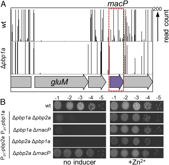
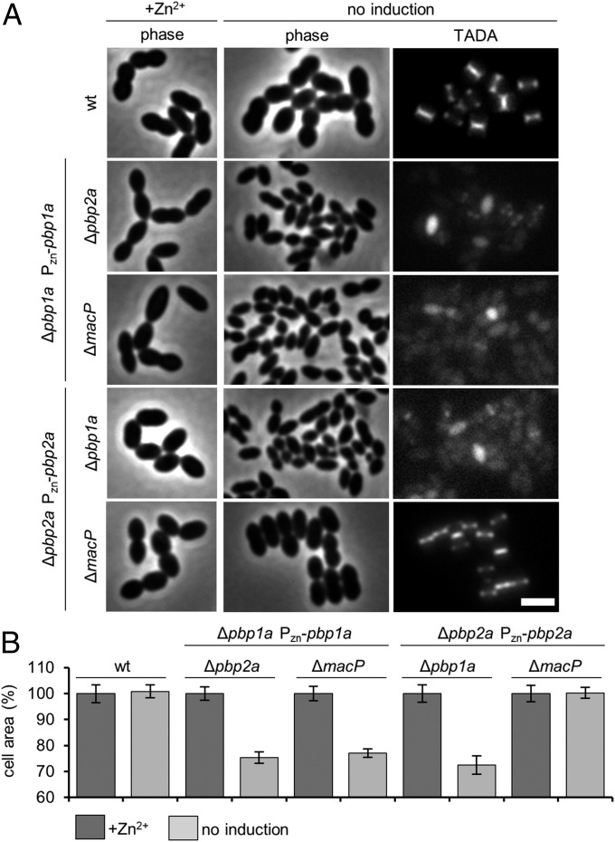
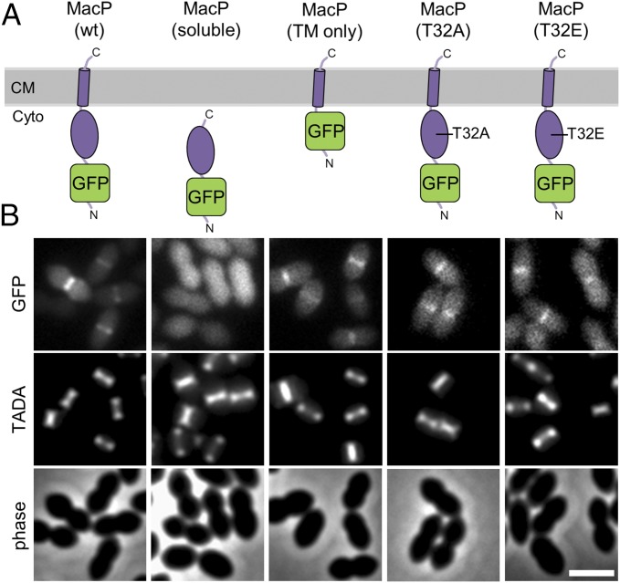
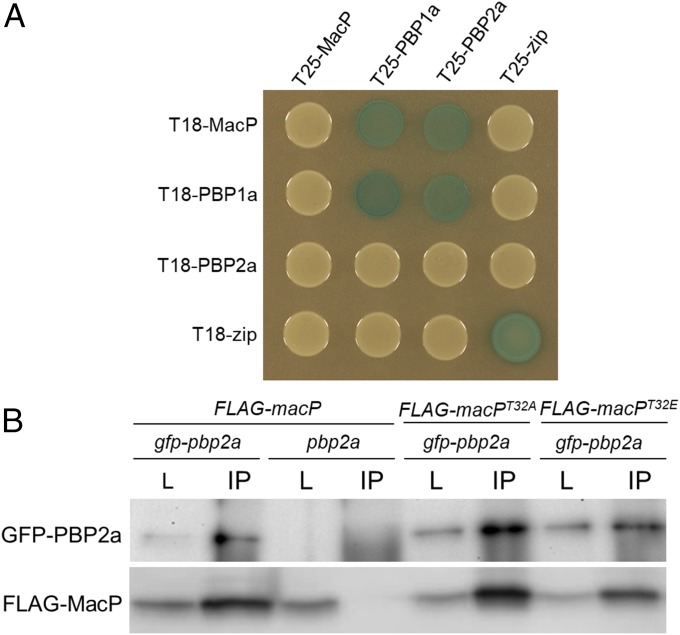
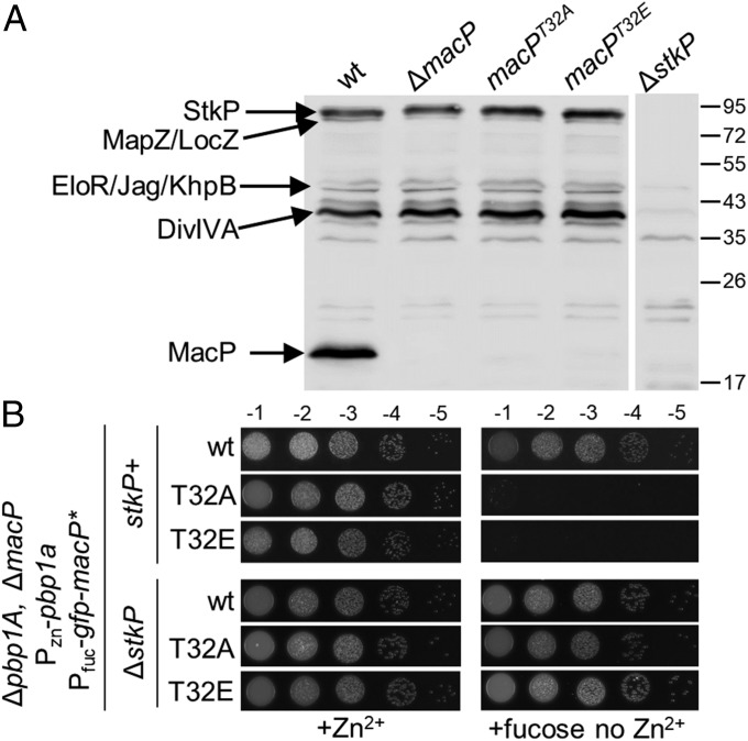
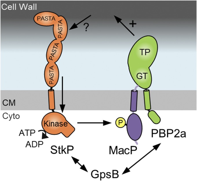
Most bacterial cells are surrounded by an essential cell wall composed of the net-like heteropolymer peptidoglycan (PG). Growth and division of bacteria are intimately linked to the expansion of the PG meshwork and the construction of a cell wall septum that separates the nascent daughter cells. Class A penicillin-binding proteins (aPBPs) are a major family of PG synthases that build the wall matrix. Given their central role in cell wall assembly and importance as drug targets, surprisingly little is known about how the activity of aPBPs is controlled to properly coordinate cell growth and division. Here, we report the identification of MacP (SPD_0876) as a membrane-anchored cofactor of PBP2a, an aPBP synthase of the Gram-positive pathogen Streptococcus pneumoniae We show that MacP localizes to the division site of S. pneumoniae, forms a complex with PBP2a, and is required for the in vivo activity of the synthase. Importantly, MacP was also found to be a substrate for the kinase StkP, a global cell cycle regulator. Although StkP has been implicated in controlling the balance between the elongation and septation modes of cell wall synthesis, none of its substrates are known to modulate PG synthetic activity. Here we show that a phosphoablative substitution in MacP that blocks StkP-mediated phosphorylation prevents PBP2a activity without affecting the MacP-PBP2a interaction. Our results thus reveal a direct connection between PG synthase function and the control of cell morphogenesis by the StkP regulatory network.

Keywords: PBP; bacterial physiology; cell division; cell wall; peptidoglycan.


Conflict of interest statement

The authors declare no conflict of interest.

Figures

Fig. 1.
Fig. 1.
macP is essential in cells lacking PBP1a. (A) Transposon insertion profiles for a select region of the chromosome for wild-type and the ∆pbp1a mutant. The height of each line in the profile represents the number of sequencing reads corresponding to a transposon insertion at the indicated genome position. Transposon insertions in the macP gene were significantly (P < 0.0002) underrepresented in the ∆pbp1a mutant relative to wild-type. Virtually no insertions were mapped to the essential gene gluM in either strain. Tn-seq output and statistical data for all libraries used in this study are shown in Fig. S1. (B) Spot dilutions of wild-type D39 ∆cps (wt) and the indicated derivatives. Cells were grown to exponential phase in the presence of 200 µM ZnCl2. The resulting cultures were normalized to an OD600 of 0.2, 10-fold serially diluted, and spotted (5 µL) onto TSA 5%SB plates in the presence or absence of 200 µM ZnCl2. Plates were incubated at 37 °C in 5% CO2 and imaged.
Fig. 2.
Fig. 2.
Loss of essential aPBP activity results in a lethal cell size defect. (A) Representative phase-contrast and fluorescence images of the indicated strains. Midexponential phase cultures were diluted to an OD600 of 0.025 in the presence or absence of 200 µM ZnCl2 inducer. Cells were grown at 37 °C in 5% CO2 for 5 h 45 min. Cells were labeled with TADA for 15 min before imaging on 2% agarose pads. n ≥ 3. (Scale bar, 3 µm.) Additional controls and time-lapse movies can be found in Fig. S2A and Movie S1, respectively. (B) Cells depleted for aPBP activity show similar reductions in cell area. Strains were grown and imaged as described in A. Cell areas were calculated from cell meshes generated in MicrobeTracker (40): 750 cells were measured in total for each condition, 250 per experiment n = 3. Error bars show SD around the mean of each experiment.
Fig. 3.
Fig. 3.
MacP localizes to sites of new PG synthesis at midcell. (A) Schematic representation of GFP–MacP and truncations used for domain analysis. The domain architecture and membrane topology of MacP are based on protease protection data and in silico predictions shown in Fig. S3. Only the full-length GFP–MacP fusion was functional as assayed by the ability to complement ∆pbp1amacP synthetic lethality (Figs. S4 and S5). The MacP soluble domain was taken as residues 1–85 and the TM segment residues 86–104. Immunoblot analysis of fusion proteins is shown in Fig. S5A. (B) Representative fluorescent and phase-contrast images of the fusions shown in A. GFP–MacP was enriched at sites of new PG synthesis at midcell. The fusions shown in A were expressed from a fucose-inducible promoter in a ΔmacP mutant inserted at the native locus. Strains were grown in Todd Hewitt broth containing 0.5% yeast extract (THY) in the presence of 0.4% fucose at 37 °C in 5% CO2 to an OD600 ∼ 0.2. Cells were labeled with TADA for 15 min before imaging on 2% agarose pads. n = 3. (Scale bar, 2 µm.)
Fig. 4.
Fig. 4.
MacP interacts with both PBP2a and PBP1a. (A) BACTH interactions between MacP and aPBP enzymes. E. coli strain BTH101 (Δcya) expressing protein fusions to domains (T25 and T18) of adenylate cyclase. Strains were grown to stationary phase and 5 µL spotted on LB agar plates containing X-gal, incubated at 30 °C, and imaged. The “zip” fusions are to a leucine zipper domain and serve as both positive and negative controls. Additional controls are provided in Fig. S7. n = 3. (B) Coimmunoprecipitation of GFP–PBP2a and a functional FLAG–MacP fusion. Each fusion was expressed from the Pzn promoter using 400 µM ZnCl2. Digitonin-solubilized membrane preparations from the indicated strains were incubated with anti-GFP resin, washed, and eluted in sample buffer. Immunoblots show matched samples of solubilized membrane fractions (L) and immunoprecipitate (IP). IP samples are 20× concentrated relative to load. Fractions were probed with anti-GFP and anti-FLAG antibodies. Representative blots are shown, n = 3. Evidence of FLAG–MacP functionality, antibody specificity, additional controls, and full immunoblot analysis are provided in Fig. S8.
Fig. 5.
Fig. 5.
The in vivo function of MacP requires phosphorylation by StkP. (A) Antiphospho-threonine immunoblot analysis of whole-cell lysates from the indicated strains. Phosphorylated StkP and its substrates are indicated based on work from previous studies (18, 19, 29). The positions of protein markers are indicated in kiloDaltons. (B) GFP–MacP(T32A)/(T32E) do not support growth of ∆pbp1A cells depleted of MacP. The indicated strains were grown to exponential phase in the presence of 200 µM ZnCl2, normalized to an OD600 of 0.2, and 5 µL of serially dilutions were spotted onto TSAII 5%SB plates in the presence of 0.2% fucose or 200 µM ZnCl2. Plates were incubated at 37 °C in 5% CO2 and imaged. Microscopy images of lethal cell size phenotype of MacP T32A and T32E on depletion of PBP1a are shown in Fig. S5C. Growth curves of each strain are provided in Fig. S5D.
Fig. 6.
Fig. 6.
StkP-dependent phosphorylation of MacP is required for PBP2a activity in vivo. Schematic model of StkP-dependent regulation of PBP2A. The protein shapes and domains are adapted from published structural information (PDB ID codes 3PY9 and 1O6Y) and data from Fig. S3. StkP senses an unknown signal through its extracellular PASTA repeats triggering its kinase domain to phosphorylate MacP at T32. Phosphorylated MacP in complex with PBP2a then stimulates PBP2a-dependent cell wall synthesis (+). GpsB interacts with StkP and is required to maintain full kinase activity and is also required for PBP2a activity. GpsB data adapted from ref. . CM, cell membrane; Cyto, cytoplasm.

References

    1. Meeske AJ, et al. SEDS proteins are a widespread family of bacterial cell wall polymerases. Nature. 2016;537:634–638. - PMC - PubMed
    1. Cho H, et al. Bacterial cell wall biogenesis is mediated by SEDS and PBP polymerase families functioning semi-autonomously. Nat Microbiol. 2016;1:16172. - PMC - PubMed
    1. Typas A, Banzhaf M, Gross CA, Vollmer W. From the regulation of peptidoglycan synthesis to bacterial growth and morphology. Nat Rev Microbiol. 2011;10:123–136. - PMC - PubMed
    1. Massidda O, Nováková L, Vollmer W. From models to pathogens: How much have we learned about Streptococcus pneumoniae cell division? Environ Microbiol. 2013;15:3133–3157. - PubMed
    1. Tsui HT, et al. Pbp2x localizes separately from Pbp2b and other peptidoglycan synthesis proteins during later stages of cell division of Streptococcus pneumoniae D39. Mol Microbiol. 2014;94:21–40. - PMC - PubMed

Publication types

MeSH terms