. 2018 Feb 27;9(1):e00130-18.
doi: 10.1128/mBio.00130-18.

Calcium-Dependent Protein Kinase 5 Is Required for Release of Egress-Specific Organelles in Plasmodium falciparum

Affiliations

Calcium-Dependent Protein Kinase 5 Is Required for Release of Egress-Specific Organelles in Plasmodium falciparum

Sabrina Absalon et al. mBio. .

Abstract

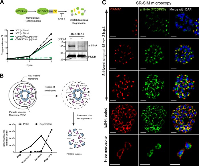
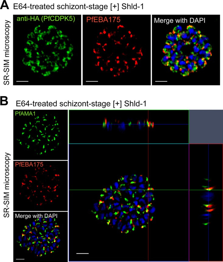
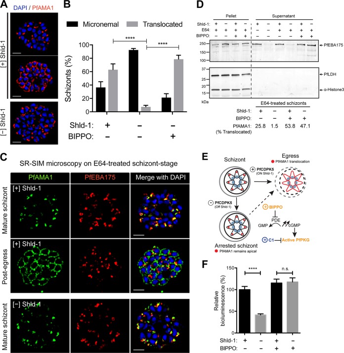
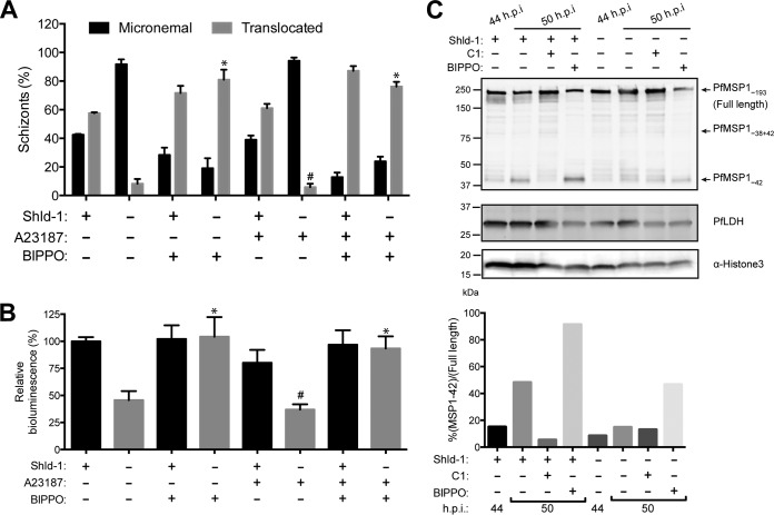
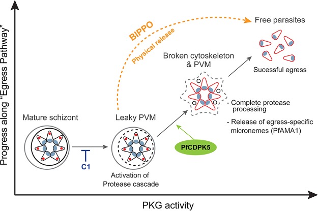
The human malaria parasite Plasmodium falciparum requires efficient egress out of an infected red blood cell for pathogenesis. This egress event is highly coordinated and is mediated by several signaling proteins, including the plant-like Pfalciparum calcium-dependent protein kinase 5 (PfCDPK5). Knockdown of PfCDPK5 results in an egress block where parasites are trapped inside their host cells. The mechanism of this PfCDPK5-dependent block, however, remains unknown. Here, we show that PfCDPK5 colocalizes with a specialized set of parasite organelles known as micronemes and is required for their discharge, implicating failure of this step as the cause of the egress defect in PfCDPK5-deficient parasites. Furthermore, we show that PfCDPK5 cooperates with the Pfalciparum cGMP-dependent kinase (PfPKG) to fully activate the protease cascade critical for parasite egress. The PfCDPK5-dependent arrest can be overcome by hyperactivation of PfPKG or by physical disruption of the arrested parasite, and we show that both treatments facilitate the release of the micronemes required for egress. Our results define the molecular mechanism of PfCDPK5 function and elucidate the complex signaling pathway of parasite egress.IMPORTANCE The signs and symptoms of clinical malaria result from the replication of parasites in human blood. Efficient egress of the malaria parasite Plasmodium falciparum out of an infected red blood cell is critical for pathogenesis. The Pfalciparum calcium-dependent protein kinase 5 (PfCDPK5) is essential for parasite egress. Following PfCDPK5 knockdown, parasites remain trapped inside their host cell and do not egress, but the mechanism for this block remains unknown. We show that PfCDPK5 colocalizes with parasite organelles known as micronemes. We demonstrate that PfCDPK5 is critical for the discharge of these micronemes and that failure of this step is the molecular mechanism of the parasite egress arrest. We also show that hyperactivation of the cGMP-dependent kinase PKG can overcome this arrest. Our data suggest that small molecules that inhibit the egress signaling pathway could be effective antimalarial therapeutics.

Keywords: Plasmodium falciparum; calcium-dependent protein kinase; egress; malaria; microneme.


Figures

FIG 1
FIG 1
Characterization of 3D7-PfCDPK5DDKnL parasites and PfCDPK5 localization. (A, top) Schematic of 3D7-PfCDPK5DDKnL regulation. This parasite strain has two genetic modifications. The destabilizing domain (DD) is fused to the carboxy terminus of PfCDPK5, and the KnL reporter gene is integrated into the genome. The PfCDPK5 fusion protein is stabilized in the presence of Shld-1 and degraded in the absence of Shld-1. (Bottom left) Replication curves of 3D7-PfCDPK5DDKnL and control 3D7 parasites cultured with Shld-1 ([+] Shld-1) and without Shld-1 ([−] Shld-1). Values are means ± standard deviations (SD) (error bars) (n = 3). (Bottom right) Immunoblot of schizont-stage lysates probed with anti-HA (recognizes the epitope tag of PfCDPK5-3HA-DD) and anti-PfLDH (loading control). The positions of molecular mass markers (in kilodaltons) are indicated to the left of the blot. h.p.i., hours post-invasion. (B) Schematic of KnL reporter protein release from 3D7-PfCDPK5DDKnL parasites. The graph shows bioluminescent activity in supernatant and parasite pellets from ring, trophozoite, schizont, and reinvaded parasites. Values are means ± SD (n = 3). (C) Schizonts from [+] Shld-1 3D7-PfCDPK5DDKnL parasites were fixed, probed with anti-HA and anti-PfAMA1 antibodies, and visualized by SR-SIM. Bars, 2 µm.
FIG 2
FIG 2
Localization of PfEBA175, PfAMA1, and PfCDPK5. (A and B) Schizonts from [+] Shld-1 3D7-PfCDPK5DDKnL parasites were fixed, probed with anti-HA and anti-PfEBA175 (A) or anti-PfAMA1 and anti-PfEBA175 (B) antibodies, and visualized by SR-SIM. The xz plane and yz plane are shown at the top and right, respectively, for panel B. The crosshairs indicate one of the limited sites of colocalization. Bars, 2 µm.
FIG 3
FIG 3
PfAMA1 translocation and egress in 3D7-PfCDPK5DDKnL parasites. (A) SR-SIM IFA of [+] Shld-1 schizonts or [−] Shld-1 schizonts probed with anti-PfAMA1 antibodies. Representative parasites with micronemal and translocated PfAMA1 are shown. Bars, 2 µm. (B) Quantification of each localization by wide-field IFA from [+] or [−] Shld-1 schizonts and from [−] Shld-1 schizonts treated with 2 µM BIPPO shown on the graph. Values are means plus standard deviations (SD) (error bars). Three biological replicates and 100 schizonts were counted per replicate. Values that were significantly different (P < 0.0001) by one-way analysis of variance (ANOVA) are indicated by bars and four asterisks. (C) SR-SIM IFAs performed on [+] Shld-1 and [−] Shld-1 3D7-PfCDPK5DDKnL schizonts demonstrating that the localization of PfEBA175 was not different in pre- and post-“egress trigger” [+] Shld-1 schizonts or in [−] Shld-1 schizonts. (D) Immunoblot analysis of PfEBA175 processing. [+] and [−] Shld-1 schizonts were treated with (+) or without (−) BIPPO. Equivalent amounts of schizont lysate and supernatant were subjected to immunoblot analysis with anti-PfEBA175, anti-PfLDH (loading control), and anti-H3 (α-Histone 3) (additional loading control) antibodies. In one set of supernatant samples, the schizonts were allowed to egress naturally without E64. The percentage of schizonts with translocated PfAMA1 is displayed below the immunoblot (similar to panel B, 100 schizonts counted per condition). (E) Schematic showing the effects of Shld-1, BIPPO, and compound 1 (C1) on PfAMA1 translocation. PDE, phosphodiesterase. (F) Quantification of bioluminescence in supernatant from [+] or [−] Shld-1 parasites at 50 h.p.i. with or without BIPPO treatment 90 min prior to measurement. Three biological replicates each done with technical triplicates were performed. Values are means ± SD (error bars). Values that are significantly different (P < 0.0001) by one-way ANOVA are indicated by a bar and four asterisks. Values that are not significantly different by one-way ANOVA (n.s.) are also indicated.
FIG 4
FIG 4
Calcium and cGMP signaling pathways in 3D7-PfCDPK5DDKnL parasites. (A) PfAMA1 translocation was visualized by wide-field IFA from [+] Shld-1 or [−] Shld-1 parasites treated with 1 µM A23187 and/or 2 µM BIPPO as indicated. Three biological replicates were performed, and 100 schizonts counted per replicate. Values are means plus SD (error bars). The percentage translocated from the small-molecule-treated [−] Shld1 samples was compared to the value for the untreated [−] Shld1 condition by one-way ANOVA. Values that were significantly different (P < 0.001) by one-way ANOVA are indicated by an asterisk. Values that were not significantly different (#) by one-way ANOVA are also indicated. (B) Bioluminescence activity released in culture supernatants from the same conditions as in panel A (three biological replicates each done with technical triplicates; mean ± SD). Each of the [−] Shld1 with small-molecule treatments was compared to the untreated [−] Shld1 condition by one-way ANOVA (*, P < 0.001; #, not significantly different). (C) 44 h.p.i. schizonts from [+] or [−] Shld-1 parasites were isolated by magnetic purification and lysed immediately or incubated for an 6 additional hours with the indicated compounds prior to lysis. Protein lysates were subjected to immunoblot analysis with anti-PfMSP142, anti-PfLDH (loading control), and anti-H3 (additional loading control) antibodies. Full-length, partially processed, and fully processed PfMSP142 are labeled. The quantitative ratio of fully processed PfMSP142 relative to the unprocessed form was calculated by volumetric measurement of fluorescence intensity with the Li-Cor Odyssey CLx system.
FIG 5
FIG 5
Physically released merozoites are invasive. (A) Schematic of the method to generate viable free merozoites efficiently from [+] Shld-1 and [−] Shld-1 schizonts. 28G, 28-gauge; HCT, hematocrit. (B) Ring parasitemia from physically released [+] or [−] Shld-1 schizonts. The ring parasitemia was not different between the [+] and [−] Shld-1 conditions (n = 3; mean ± SD; no significant difference by Student’s t test). (C) Wide-field IFA of PfAMA1 localization before and after shearing from [+] and [−] Shld-1 parasites. Treatment of schizonts with 2.5 µM C1 prior to shearing prevents PfAMA1 translocation.
FIG 6
FIG 6
Model for cooperativity between PfCDPK5 and PfPKG. The progress along the parasite egress pathway is promoted by activation of PfPKG. At low levels of PfPKG activation, the protease cascade and calcium signaling pathway are initiated. PfCDPK5 activation, together with PfPKG, leads to further progression of the protease cascade and triggered release of micronemes required for parasite egress (detected by PfAMA1 translocation, shown in red). Under physiological conditions, this step requires both PfCDPK5 and PfPKG. The discharge of micronemes completes the egress process with the release of invasive merozoites. The requirement for PfCDPK5 can be bypassed by supraphysiological activation of PfPKG by either BIPPO or physical disruption. PVM, parasitophorous vacuolar membrane.

References

    1. World Health Organization 2016. World malaria report 2015. World Health Organization, Geneva, Switzerland.
    1. Ashley EA, Dhorda M, Fairhurst RM, Amaratunga C, Lim P, Suon S, Sreng S, Anderson JM, Mao S, Sam B, Sopha C, Chuor CM, Nguon C, Sovannaroth S, Pukrittayakamee S, Jittamala P, Chotivanich K, Chutasmit K, Suchatsoonthorn C, Runcharoen R, Hien TT, Thuy-Nhien NT, Thanh NV, Phu NH, Htut Y, Han K-T, Aye KH, Mokuolu OA, Olaosebikan RR, Folaranmi OO, Mayxay M, Khanthavong M, Hongvanthong B, Newton PN, Onyamboko MA, Fanello CI, Tshefu AK, Mishra N, Valecha N, Phyo AP, Nosten F, Yi P, Tripura R, Borrmann S, Bashraheil M, Peshu J, Faiz MA, Ghose A, Hossain MA, Samad R, Rahman MR, et al. . 2014. Spread of artemisinin resistance in Plasmodium falciparum malaria. N Engl J Med 371:411–423. doi:10.1056/NEJMoa1314981. - DOI - PMC - PubMed
    1. White NJ, Pukrittayakamee S, Hien TT, Faiz MA, Mokuolu OA, Dondorp AM. 2014. Malaria. Lancet 383:723–735. doi:10.1016/S0140-6736(13)60024-0. - DOI - PubMed
    1. Miller LH, Ackerman HC, Su X-Z, Wellems TE. 2013. Malaria biology and disease pathogenesis: insights for new treatments. Nat Med 19:156–167. doi:10.1038/nm.3073. - DOI - PMC - PubMed
    1. Cowman AF, Healer J, Marapana D, Marsh K. 2016. Malaria: biology and disease. Cell 167:610–624. doi:10.1016/j.cell.2016.07.055. - DOI - PubMed

Publication types

MeSH terms

Substances

LinkOut - more resources