. 2018 Feb 27;8(1):3746.
doi: 10.1038/s41598-018-22139-6.

Influenza A viruses alter the stability and antiviral contribution of host E3-ubiquitin ligase Mdm2 during the time-course of infection

Affiliations

Influenza A viruses alter the stability and antiviral contribution of host E3-ubiquitin ligase Mdm2 during the time-course of infection

Andrés Pizzorno et al. Sci Rep. .

Abstract

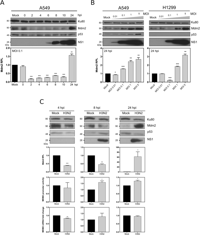
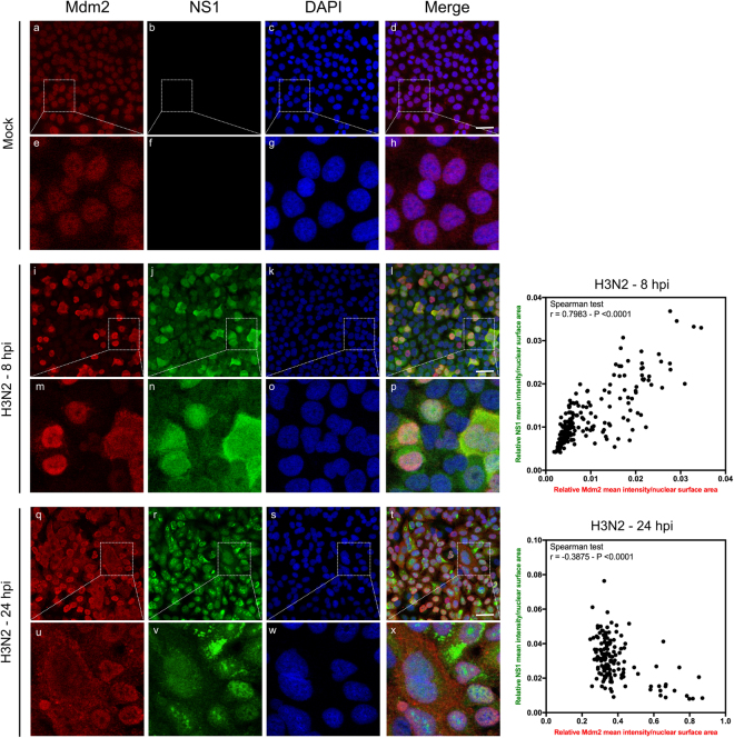
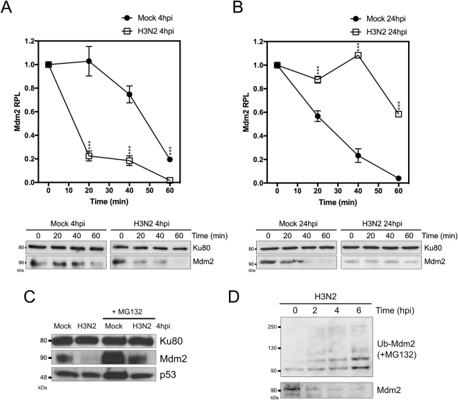
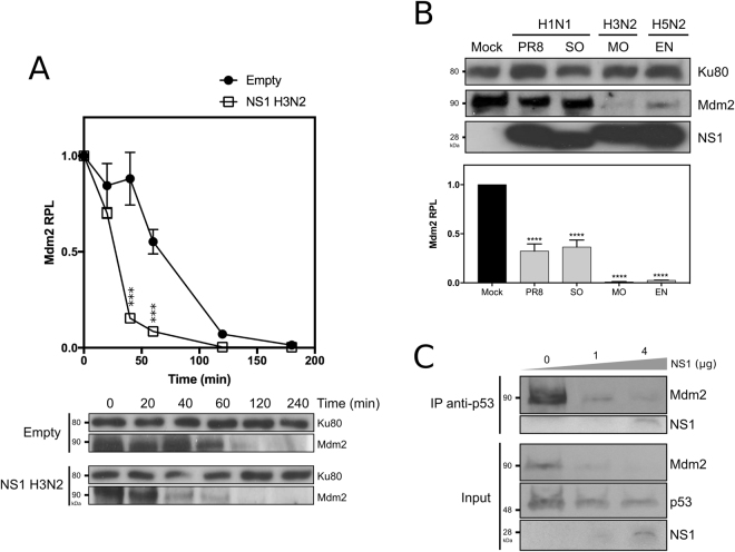
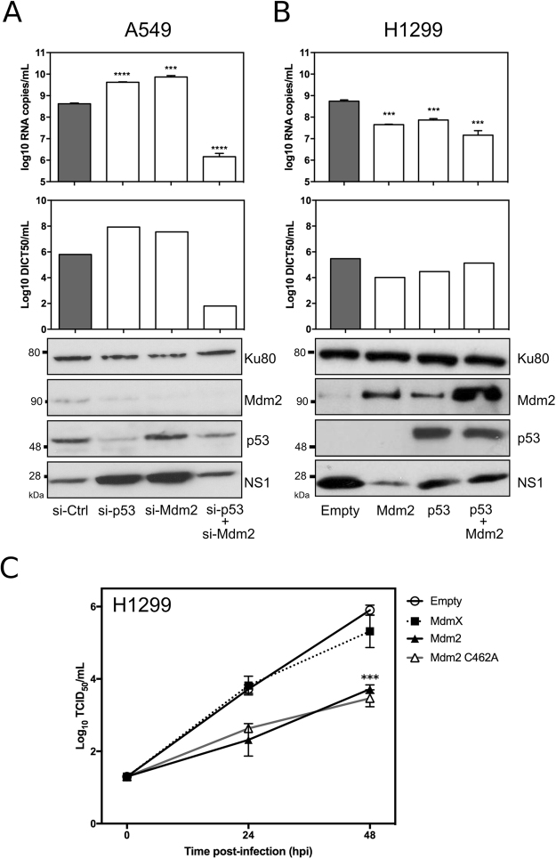
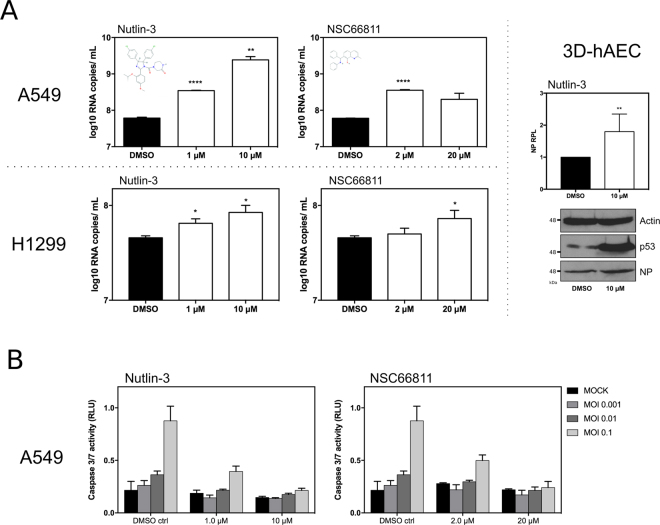
The interplay between influenza A viruses (IAV) and the p53 pathway has been reported in several studies, highlighting the antiviral contribution of p53. Here, we investigated the impact of IAV on the E3-ubiquitin ligase Mdm2, a major regulator of p53, and observed that IAV targets Mdm2, notably via its non-structural protein (NS1), therefore altering Mdm2 stability, p53/Mdm2 interaction and regulatory loop during the time-course of infection. This study also highlights a new antiviral facet of Mdm2 possibly increasing the list of its many p53-independent functions. Altogether, our work contributes to better understand the mechanisms underlining the complex interactions between IAV and the p53 pathway, for which both NS1 and Mdm2 arise as key players.


Conflict of interest statement

O.T., J.C.B. and M.R.C. are co-inventors of a patent application deposited by University of Dundee, Centre National de la Recherche Scientifique, Université Claude Bernard Lyon 1 and Hospices Civils de Lyon (FR20100059132 20101105; WO2011FR52575 20111104). The other authors declare that they have no competing interests.

Figures

Figure 1
Figure 1
Mdm2 expression is strongly impacted by IAV infection, mostly at post-transcriptional levels. (A) Human lung A549 cells were mock-infected or infected by influenza A/Moscow/10/99 (H3N2) at a MOI of 0.1 and cell lysates were analyzed by western blot for the expression of Mdm2 (SMP14 antibody), p53 and IAV NS1 at different time-points post infection. Ku80 was used as a loading control. Mdm2 relative protein levels (Mdm2 RPL) were measured by densitometry and calculated from data from three independent experiments. An asterisk indicates a significant difference compared with the results for mock-infected cells (** and *** for P-value < 0.01 and 0.001, respectively). (B) Alternatively, A549 or H1299 cells were mock-infected or infected by influenza A/Moscow/10/99 (H3N2) at different MOI and expression of Mdm2, p53 and IAV NS1 were monitored at 24 h post-infection by western blot, using the same approach. (C) A549 cells were mock-infected or infected by influenza A/Moscow/10/99 (H3N2) at a MOI of 4 or 0.1 and cell supernatants and lysates were harvested at 4, 8 or 24 hpi, respectively. Endogenous Mdm2 expression was measured at protein level by western blot, Mdm2 promoter activity was monitored using a luciferase reporter plasmid. In addition, Mdm2 mRNA expression was measured by RT-qPCR. An asterisk indicates a significant difference compared with the results for mock-infected cells (*, **, *** for P-value < 0.05, 0.01 and < 0.001, respectively).
Figure 2
Figure 2
IAV alters Mdm2 protein level and its nucleo-cytoplasmic localization during the time-course of infection. Immunofluorescence staining of Mdm2 (red) and influenza NS1 (green) in mock-infected (panels a to h) or infected with H3N2 virus at a MOI of 1 (panels i to x) was performed at different times, as indicated. Nuclei were counterstained with DAPI (blue, panels c,g,k,o,s,w). Merged fluorescent signals are presented in panels d,h,i,p,t and x. Cell details are enlarged (inset). White scale bar = 10 μm. The presented results obtained by confocal immunofluorescence microscopy are representative fields, from several repeated experiments. The relative mean nuclear intensity of NS1 and Mdm2 stainings was measured at 8 and 24 hpi with ImageJ (version 1.51 h - http://imagej.nih.gov/ij), using DAPI staining to define nuclear areas for measurements. Data collected for NS1 and Mdm2 were subject to correlation analysis.
Figure 3
Figure 3
IAV infection modulates Mdm2 stability. Stability assay in IAV-infected cells at 4 hpi (A) and 24 hpi (B). Human lung A549 cells were mock-infected or infected with influenza A/Moscow/10/99 (H3N2) with an MOI of 4 or 0.1 and analyzed at 4 and 24 hpi, respectively. Stability was assessed by monitoring relative protein levels (RPL) of Mdm2 during a 1 h period, after treatment with 50 µM cycloheximide (CHX). Mean values +/− standard deviation from three independent experiments are presented. An asterisk indicates a significant difference compared with the results for mock-infected cells (*** for P-value < 0.001). (C) Human lung A549 cells were mock-infected or infected with influenza A/Moscow/10/99 (H3N2) with an MOI of 4, in presence/absence of proteasome inhibitor MG132 (20 µM), and harvested at 4 h post-infection, and analyzed by western blot. (D) A549 cells were transfected with a plasmid expressing His-tagged Ubiquitin and then further infected 48 h post-transfection with influenza A/Moscow/10/99 (H3N2) with an MOI of 4, in presence of 20 µM MG132. Cell lysates were harvested at different time-points after infection, ubiquitinated products were separated and analyzed by western blot using a specific antibody against Mdm2. In parallel, without MG132 treatment, Mdm2 protein levels were monitored at similar time-points. When necessary, Ku80 was used as a loading control for western blot.
Figure 4
Figure 4
IAV NS1 expression contributes to IAV-induced Mdm2 destabilization, and consecutively alters Mdm2/p53 interaction. (A) Stability assay in presence of NS1. A549 cells were transfected with either an empty plasmid or a plasmid expressing NS1 from A/Moscow/10/00 (H3N2), and Mdm2 stability was evaluated at 48 h post-transfection. Stability was assessed by monitoring relative protein levels (RPL) of Mdm2 during a 1 h period, after treatment with 50 µM cycloheximide (CHX). Mean values +/− standard deviation from three independent experiments are presented. *** for P-value < 0.001. Ku80 was used as a loading control. (B) Four different recombinant IAV were generated using reverse genetics, using the same A/PuertoRico/8/34 (H1N1) genomic background, and harboring NS segment from different IAV strains; NS from A/PuertoRico/8/34 (H1N1, PR8), swine-origin A/Lyon/0969/09 (H1N, SO), A/Moscow/10/99 (H3N2, MO), A/Finch/England/20151/94 (H5N2, EN). Human lung A549 cells were mock-infected or infected with these different IAV with an MOI of 4 and cell lysates were harvested at 8 h post-infection for western blot analysis. Mdm2 relative protein levels (Mdm2 RPL) were measured by densitometry and calculated from data from three independent experiments. An asterisk indicates a significant difference compared with the results for mock-infected cells (**** for P-value < 0.0001). Ku80 was used as a loading control. (C) Impact of NS1 transient expression on the interaction between p53 and Mdm2. A549 cells were transfected with either an empty plasmid, or two quantities of plasmids expressing NS1 (1 and 4 µg NS1 H3N2). After 48 h post-transfection, cells lysates were analyzed using a co-immunoprecipitation assay using an anti-p53 polyclonal antibody.
Figure 5
Figure 5
An unexpected antiviral contribution of Mdm2 revealed by silencing/transient expression experiments. (A) Knock-down of p53 and/or Mdm2 mRNA expression in A549 cells differentially modulates levels of IAV production (Left panel). Forty-eight hours after si-RNA transfection using a control si-RNAs (si-Ctrl) or specific siRNAs (si-p53/si-Mdm2), A549 cells were infected by A/Moscow/10/99 (H3N2) at a MOI of 0.01 and the level of viral production at 24 h post-infection was assessed using three different experiments: (i) RT-qPCR (log10 RNA copies/mL, measured in three independent experiments. An asterisk indicates a significant difference compared with the results for si-Ctlr treated cells (*** and **** for respectively P-values < 0.001 and <0.0001), (ii) determination of infectious viral titers of supernatants (TCID50/mL) by endpoint titration in MDCK cells (measured in quadruplicate in 2 independent experiments), and (iii) western blot. Ku80 was used as a loading control; (B) Transient expression/co-expression of p53 and/or Mdm2 in H1299 cells decreases the level of viral production (Right panel). Forty-eight hours after transfection, H1299 cells were infected by A/Moscow/10/99 (H3N2) at a MOI of 0.01 and the level of viral production at 24 h post-infection was assessed using similar methods. An asterisk indicates a significant difference compared with the results obtained for cells transfected with the empty vector (*** and **** for respectively P-values < 0.001 and <0.0001) (C) Comparative viral kinetics in the context of transient expression of Mdm2, Mdm2 RING mutant C462A or Mdmx in H1299 cells. Forty-eight hours after transfection, H1299 cells were infected by A/Moscow/10/99 (H3N2) at a MOI of 0.01 and the level of viral production at 24 h and 48 h post-infection was assessed using determination of infectious titers of supernatants (log10 TCID50/mL) by endpoint titration in MDCK cells (measured in quadruplicate in 2 independent experiments). An asterisk indicates a significant difference compared with the results obtained for cells transfected with the empty vector (*** for P-value < 0.001).
Figure 6
Figure 6
An unexpected antiviral contribution of Mdm2 revealed by small-molecules Mdm2 antagonists. (A) Impact of small molecule Mdm2 antagonists on viral production and IAV-induced apoptosis. Human lung epithelial A549 or H1299 cells were infected by influenza virus A/Moscow/10/99 (H3N2) at a MOI of 0.001, in presence of DMSO or small molecules Mdm2 antagonists (Nutlin-3 or NSC 66811) at different concentrations. The level of viral production at 48 h post-infection was evaluated by RT-qPCR (log10 RNA copies/mL measured in three independent experiments). An asterisk indicates a significant difference compared with the results for DMSO treated cells (*, **, **** for P-values < 0.01, <0.05 and <0.0001, respectively). 3D-Human airway epithelia (3D-hAEC), which are constituted by human primary respiratory epithelial cells cultivated at the air-liquid interface, were treated in basal medium with Nutlin-3 (10 μM) or DMSO and then infected by influenza virus A/Moscow/10/99 (H3N2) at a MOI of 0.1. Cell lysates were analyzed at 48 hours post-infection by western blot for the expression of IAV NP and p53. Actin was used as a loading control. IAV NP relative protein levels (NP RPL) were measured by densitometry and calculated from data from two independent experiments. (B) The impact of small molecule antagonists on IAV-induced caspase3/7 activity at 48 h post-infection in A549 cells was monitored using a luciferase reporter assay, using three different MOI (0.1, 0.01 and 0.001).

References

    1. Paules C, Subbarao K. Influenza. Lancet Lond Engl. 2017 - PubMed
    1. Neumann G, Kawaoka Y. The first influenza pandemic of the new millennium. Influenza Other Respir Viruses. 2011;5:157–166. doi: 10.1111/j.1750-2659.2011.00231.x. - DOI - PMC - PubMed
    1. Fukuyama S, Kawaoka Y. The pathogenesis of influenza virus infections: the contributions of virus and host factors. Curr Opin Immunol. 2011;23:481–486. doi: 10.1016/j.coi.2011.07.016. - DOI - PMC - PubMed
    1. Stertz S, Shaw ML. Uncovering the global host cell requirements for influenza virus replication via RNAi screening. Microbes Infect Inst Pasteur. 2011;13:516–525. doi: 10.1016/j.micinf.2011.01.012. - DOI - PMC - PubMed
    1. König R, et al. Human host factors required for influenza virus replication. Nature. 2010;463:813–817. doi: 10.1038/nature08699. - DOI - PMC - PubMed

MeSH terms

Substances